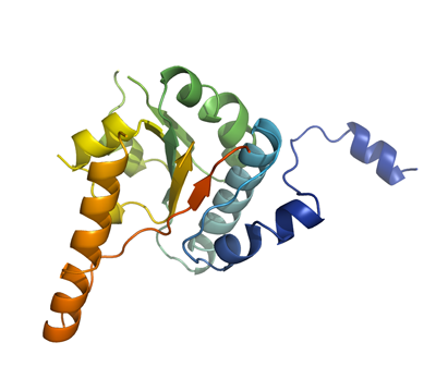
MDA5
PDB:3B6E
Revision
Revision Type:created
Revised by:created
Revision Date:created
Entry Clone Accession:BC111750
Entry Clone Source:Mammalian Gene Collection
SGC Clone Accession:Tag:N-terminal hexahistidine tag with integrated TEV protease cleavage site: mhhhhhhssgvdlgtenlyfq*sm
Host:E.coli BL21-Gold(DE3)pRARE2, where BL21-Gold(DE3) cells (Stratagene) have been transformed with pRARE2 originating from the Rosetta2 strain (Novagen). The pRARE2 plasmid supplies tRNAs for rare codons.
Construct
Prelude:Sequence:mhhhhhhssgvdlgtenlyfq*smNSNMGSDSGTMGSDSDEENVAARASPEPELQLRPYQMEVAQPALEGKNIIICLPTGSGKTRVAVYIAKDHLDKKKKASEPGKVIVLVNKVLLVEQLFRKEFQPFLKKWYRVIGLSGDTQLKISFPEVVKSCDIIISTAQILENSLLNLENGEDAGVQLSDFSLIIIDECHHTNKEAVYNNIMRHYLMQKLKNNRLKKENKPVIPLPQILGLTAS
Vector:pNIC-Bsa4
Growth
Medium:Antibiotics:Procedure:Native protein
Cells from a glycerol stock were streaked onto LB-agar plates. 5-10 colonies were used to inoculate 2 x 20 ml TB supplemented with 8 g/l glycerol, 100 µg/ml kanamycin and 34 µg/ml chloramphenicol and the cells were grown at 30 ºC overnight. The overnight cultures were used to inoculate 2 x 1.5 l TB supplemented with 8 g/l glycerol, 50 µg/ml kanamycin, 34 µg/ml chloramphenicol and approximately 200 µl PPG P2,000 81380 anti-foam solution (Fluka). The cultures were grown in a LEX bioreactor system (Harbinger Biotechnology) at 37 ºC until OD600 reached ~1.3. The cultures were down-tempered to 18 ºC over a period of 1 hour before target expression was induced by addition of 0.5 mM IPTG. Expression was allowed to continue overnight and cells were harvested the following morning by centrifugation (5,500 x g, 15 min, 4 ºC). The resulting cell pellet (81.2 g wet cell weight) was resuspended in lysis buffer (1 ml/g cell pellet), supplemented with 1 tablet of Complete EDTA-free protease inhibitor (Roche Applied Science). The cell suspension was stored at -80 ºC.
SeMet labeled protein
Cells from a glycerol stock were streaked onto LB-agar plates. 5-10 colonies were used to inoculate 2 x 40 ml LB supplemented with 100 µg/ml kanamycin and 34 μg/ml chloramphenicol and the cells were grown at 30 ºC overnight. 60 ml of the overnight culture were used to inoculate 6 flasks with 1.5 l minimal medium (without amino acids) supplemented with 50 µg/ml kanamycin, 34 μg/ml chloramphenicol and approximately 250 µl PPG P2,000 81380 anti-foam solution (Fluka) per bottle. The cultures were grown in a LEX bioreactor system (Harbinger Biotechnology) at 37 ºC until OD600 reached ~1. The flasks were down-tempered to 18 ºC and after 30 minutes, amino acids were added (Van Duyne, G. D., J. Mol. Biol. 229, 105-124 (1993)). About one hour later, expression of target protein was induced by addition of 0.5 mM IPTG. The expression was allowed to continue at 18 ºC overnight. Cells were harvested the following morning by centrifugation (5,000 x g, 13 min, 4 ºC). The resulting cell pellet (54 g from 9 liter culture) was resuspended in lysis buffer (2 ml/g cell pellet) supplemented with two tablets of Complete EDTA-free protease inhibitor (Roche Applied Science) and stored at -80 ºC. Selenomethionine enriched protein was grown in minimal medium containing: 25 mM Na2HPO4, 25 mM KH2PO4, 50 mM NH4Cl, 5 mM Na2SO4, 0.4% (w/v) glucose, 2 mM MgSO4, 0.1 mM CaCl2, 1.0 µM MnCl2, 10 µM FeSO4, pH ~7. Mix of amino acids added (per liter culture): 100 mg each of lysine, threonine, phenylalanine and 50 mg each of leucine, isoleucine, valine, L(+)-selenomethionine.
Purification
ProcedureColumns
IMAC 1: Ni-charged 1 ml HiTrap Chelating HP (GE Healthcare)
IMAC 2: Ni-charged 1 ml HisTrap FF crude (GE Healthcare)
Gel filtration column: HiLoad 16/60 Superdex 75 Prep Grade (GE Healthcare)
Procedure
Purification of the native protein was performed as a two step process on an ÄKTAxpress system (GE Healthcare). Prior to purification, columns were equilibrated with IMAC wash1 buffer and gel filtration buffer, respectively. The filtered lysate was loaded onto the Ni-charged HiTrap Chelating column and washed with IMAC wash1 buffer followed by IMAC wash2 buffer. Bound protein was eluted from the IMAC column with IMAC elution buffer and automatically loaded onto the gel filtration column. Fractions containing the target protein were pooled and fresh TCEP was added to a final concentration of 2 mM. The protein was subsequently concentrated using an Amicon Ultra-15 centrifugal filter device, 10,000 NMWL (Millipore) to 27.2 mg/ml in a volume of 0.3 ml. The samples were stored at -80 ºC. Purification of the SeMet enriched protein was performed in basically the same way. The filtered lysate was purified by IMAC and gel filtration chromatography followed by a concentration step. The final SeMet-protein concentration was 33.5 mg/ml in a volume of 1.5 ml.
Tag removal
The N-terminal histidine tag was proteolytically removed by incubating the target protein (27.2 mg/ml in 0.3 ml) with His-tagged TEV protease at a molar ratio of 50:1 in 4 ºC overnight. The proteolytic reaction went to completion, as judged by SDS-PAGE. The reaction mixture was diluted with GF buffer to reduce the TCEP concentration. The target protein was subsequently purified from tag and protease by passing it over a Ni-charged HisTrap FF crude column (GE Healthcare) pre-equilibrated with GF buffer. The cleaved protein was eluted with IMAC wash2 buffer. The protein was concentrated and the buffer changed to GF buffer (with 2 mM TCEP) using an Amicon Ultra-15 centrifugal filter device, 10,000 NMWL (Millipore). The concentration was measured to 21.9 mg/ml in a volume of 0.35 ml. The identity of the protein was confirmed by mass spectrometry. The histidine tag removal of SeMet labeled protein was achieved in basically the same way as described for the native protein. Purified SeMet labeled protein (33.5 mg/ml) was incubated with TEV protease at 4 ºC overnight, passed over a Ni-charged HisTrap FF crude column and concentrated to a final concentration of 48.1 mg/ml in 0.8 ml.
Extraction
ProcedureThe cell suspension was quickly thawed in water and 4000 U Benzonase (Merck) were added. Cells were disrupted by sonication (VibraCell, Sonics) at 70% amplitude for 3 min effective time (puls 4s on, 4s off) and cell debris was removed by centrifugation (49,000 x
g, 30 min, 4 ºC). The supernatant was decanted and filtered through a 0.45 µm flask filter. The SeMet pellet was basically treated in the same way.
Concentration:LigandMassSpec:Crystallization:Native crystals were obtained by the sitting drop vapour diffusion method in a 96-well plate. 0.2 µl of the protein solution (diluted to 19.9 mg/ml) including 20 mM ADP and 10 mM MgCl2 was mixed with 0.1 µl of well solution consisting of 0.1 M Tris pH 8.0 and 1.2 M sodium-citrate. The plates were incubated at 20 ºC and one pyramidal crystal was obtained after one day and continued to grow for two more weeks. The crystal was quickly transferred to cryo solution consisting of well solution complemented with 20% glycerol and flash frozen in liquid nitrogen. SeMet crystals were produced in the same way by mixing 0.2 µl of the SeMet-protein solution (diluted with GF buffer to 20 mg/ml) including 20mM ADP and 10mM MgCl2 with 0.1 µl of well solution consisting of 0.1 M bis-Tris-Propane pH 7.5, and 65% Tacsimate. The plate was incubated at 20 ºC and crystals were obtained after one day and continued to grow for one more week. The crystals were quickly transferred to cryo solution consisting of well solution complemented with 20% glycerol and flash frozen in liquid nitrogen.
NMR Spectroscopy:Data Collection:Diffraction data up to 2.2 Å resolution was collected at BESSY (BL14-1).
Data Processing:The structure was solved by SAD using Seleno-methionine labeled protein crystals. The space group was P6522 with cell dimensions a=72.8 Å, b=72.8 Å, c=182.3 Å. The asymmetric unit contains one monomer. The obtained model was thereafter used as an initial model for refinement with native data to a resolution of 1.6 Å. The same space group P6522 with cell dimensions a=72.8 Å, b=72.8 Å, c=182.8 Å. One monomer was located in the asymmetric unit. Automatic model building using Arp/warp was performed; Refmac5 was used for refinement and Coot for model building. Data in the interval 20.0-1.60 Å resolution was used and at the end of the refinement the R values were: R= 17.9% and Rfree= 20.4%. Coordinates for the crystal structure were deposited in the Protein Data Bank, accession code 3B6E.