THTPA
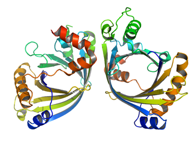
PDB:3BHD
Revision
Revision Type:created
Revised by:created
Revision Date:created
Entry Clone Accession:gi|13236577
Entry Clone Source:Mammalian Gene Collection
SGC Clone Accession:THTPAA-k021
Tag:N-terminal hexahistidine tag including a thrombin protease cleavage site: mgsshhhhhhssglvprg*s(m)
Host:E.coli BL21-Gold(DE3)pG-Tf2, denotes BL21-Gold(DE3) cells from Stratagene transformed with the chaperone coding plasmid pG-Tf2 (TaKaRa).
Construct
Prelude:Sequence:mgsshhhhhhssglvprg*sMAQGLIEVERKFLPGPGTEERLQELGGTLEYRVTFRDTYYDTPELSLMQADHWLRRREDSGWELKCPGAAGVLGPHTEYKELTAEPTIVAQLCKVLRADGLGAGDVAAVLGPLGLQEVASFVTKRSAWKLVLLGADEEEPQLRVDLDTADFGYAVGEVEALVHEEAEVPTALEKIHRLSSMLGVPAQETAPAKLIVYLQRFRPQDYQRLLEVNSS
Vector:pET28-LIC
Growth
Medium:Selenomethionine enriched protein was grown in minimal medium containing : 25 mM Na
2HPO
4, 25 mM KH
2PO
4, 50 mM NH
4Cl, 5 mM Na
2SO
4, 0.4% (w/v) glucose, 2 mM MgSO
4, 0.1 mM CaCl
2, 0.5 µM MnCl
2, 10 µM FeSO
4, pH ~7. Mix of amino acids added (per liter culture): 100 mg each of lysine, threonine, phenylalanine and 50 mg each of leucine, isoleucine, valine, L(+)-selenomethionine.
Antibiotics:Procedure:Cells from glycerol stock were grown in 100 ml SOB supplemented with 50 µg/ml kanamycin at 30 ºC overnight. 90 ml of the overnight culture were used to inoculate 12 flasks with 0.75 l minimal medium (without amino acids) supplemented with 50 µg/ml kanamycin and approximately 50 µl BREOX FMT 30 anti-foam solution (Cognis Performance Chemicals UK Ltd) per flask. The cultures were grown in TunAir flasks at 37 ºC until OD600 reached ~0.5. The flasks were down-tempered to 18 ºC, amino acids were added (Van Duyne, G. D.,
J. Mol. Biol. 229, 105-124 (1993)) and expression of chaperones was induced by addition of 2 ng/ml tetracycline. 1.5 hours later, expression of target protein was induced by addition of 0.5 mM IPTG. The expression was allowed to continue at 18 ºC overnight. Cells were harvested the following morning by centrifugation (3,500 x
g, 10 min, 4 ºC). The resulting cell pellet (51 g from 9 liter culture) was stored at -80 ºC.
Purification
ProcedureColumns
IMAC: Ni-charged 1 ml HiTrap Chelating HP (GE Healthcare)
Gel filtration column: HiLoad 16/60 Superdex 75 Prep Grade (GE Healthcare)
IEX 1: Mono S 5/50 GL (GE Healthcare)
IEX 2: Mono Q 5/50 GL (GE Healthcare)
Procedure
Purification of the SeMet-labeled protein was performed as a two step process using a peristaltic pump and an ÄKTApurifier system (GE Healthcare). Prior to purification, columns were equilibrated with IMAC wash1 buffer and gel filtration buffer, respectively. The filtered lysate was loaded onto the Ni-charged HiTrap Chelating column and washed with IMAC wash1 buffer followed by IMAC wash2 buffer. Bound protein was eluted from the IMAC column with IMAC elution buffer, concentrated to 2.5 ml and loaded onto the gel filtration column. Fractions containing the target protein were pooled and subsequently concentrated using a Vivaspin 20 centrifugal filter device, 10,000 NMWL (Sartorius). The concentration was measured to 18.2 mg/ml in a volume of 0.46 ml and stored at -80 ºC.
Tag removal
The N-terminal histidine tag was proteolytically removed by incubating the target protein (18.2 mg/ml in 0.46 ml) with thrombin (10 U/mg protein) at 4 ºC overnight. Prior to purification, columns were equilibrated with Mono S buffer A and Mono Q buffer A, respectively. After incubation, the cleaved protein sample was diluted to 5 ml in Mono S buffer A, filtered through a 0.45 µm filter and passed over a Mono S column. The flow through was collected and the sample diluted with Mono Q buffer A. The pH was then adjusted to 8.0 by addition of 1 M Tris pH 8.0, and loaded onto a Mono Q column. The column was washed with Mono Q buffer A before eluting the protein with a gradient of Mono Q buffer B. The cleaved protein was concentrated and the buffer was changed to 20 mM HEPES, 100 mM NaCl, 2 mM TCEP, pH 7.5 using a Vivaspin 20 centrifugal filter device, 10,000 MWCO (Sartorius). The final protein concentration was determined to 10.3 mg/ml in a volume of 0.15 ml. The identity of the protein was confirmed by mass spectrometry.
Extraction
ProcedureThe cell pellet was quickly thawed and resuspended in lysis buffer (1ml/g cell pellet) supplemented with one tablet of Complete EDTA-free protease inhibitor (Roche Applied Science), 2000 U Benzonase (Merck) and a knife edge of lysozyme (Sigma). Cells were disrupted by sonication (VibraCell, Sonics) at 70% amplitude for 3 min effective time (puls 4s on, 4s off) and cell debris was removed by centrifugation (49,000 x
g, 30 min, 4 ºC). The supernatant was decanted and filtered through a 0.45 µm flask filter.
Concentration:LigandMassSpec:Crystallization:SeMet crystals were obtained by the hanging drop vapour diffusion method in a 24-well plate containing 500 µl well solution. 0.8 µl of the protein solution (10.3 mg/ml) including 3 mM sodium tungstenate, was mixed with 0.8 µl of well solution consisting of 0.05 M citric acid, pH 3.8, 0.15 M ammonium sulfate. The plate was incubated at 4 ºC and one thick rod crystal appeared after 12 days, growing from a spherulite. The crystal was briefly transferred to cryo solution containing 0.05 M citric acid, pH 3.8, 0.3 M AmSO4, 0.3 M NaCl, 30% Glycerol and flash frozen in liquid nitrogen.
NMR Spectroscopy:Data Collection:Data to 1.5 Å resolution was collected at ESRF beamline ID29. A single crystal was used to collect 360º degrees oscillation range at peak, remote and inflection wavelengths for selenium.
Data Processing:Data was processed with XDS and solved by SAD using Shelx. The space group was P21 with cell dimensions a=60.77 Å, b=46.81 Å, c=71.87 Å, α=90.00, β=113.97, γ=90.00 and 2 molecules in the asymmetric unit. Automatic model building using Arp/warp was performed; Refmac5 was used for refinement and Coot for model building. Data in the interval 20.0-1.50 Å resolution was used during refinement. Final values for R and Rfree were 17.6% and 20.1%, respectively. Coordinates for the crystal structure were deposited in the Protein Data Bank, accession code 3BHD.