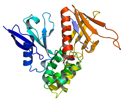
EPB41L3
PDB:3BIN
Revision
Revision Type:created
Revised by:created
Revision Date:created
Entry Clone Accession:BC006141
Entry Clone Source:Mammalian Gene Collection
SGC Clone Accession:Tag:N-terminal hexahistidine tag with integrated TEV protease cleavage site: mhhhhhhssgvdlgtenlyfq*sm
Host:E.coli BL21(DE3) (Novagen)
Construct
Prelude:Sequence:mhhhhhhssgvdlgtenlyfq*smKPKSMQCKVILLDGSEYTCDVEKRSRGQVLFDKVCEHLNLLEKDYFGLTYRDAENQKNWLDPAKEIKKQVRSGAWHFSFNVKFYPPDPAQLSEDITRYYLCLQLRDDIVSGRLPCSFVTLALLGSYTVQSELGDYDPDECGSDYISEFRFAPNHTKELEDKVIELHKSHRGMTPAEAEMHFLENAKKLSMYGVDLHHAKDSEGVEIMLGVCASGLLIYRDRLRINRFAWPKVLKISYKRNNFYIKIRPGEFEQFESTIGFKLPNHRAAKRLWKVCVEHHTFFRLLLPEAPPK
Vector:pNIC-Bsa4
Growth
Medium:Antibiotics:Procedure:Cells from a glycerol stock were grown in 20 ml TB supplemented with 8 g/l glycerol and 100 µg/ml kanamycin at 30 ºC overnight. The overnight culture (20 ml) was used to inoculate 1.5 l TB supplemented with 8 g/l glycerol, 50 µg/ml kanamycin and approximately 100 µl BREOX FMT 30 anti-foam solution (Cognis Performance Chemicals UK Ltd). The culture was grown in a LEX bioreactor system (Harbinger Biotechnology) at 37 ºC until OD600 reached ~1.7. The culture was down-tempered to 18 ºC over a period of 1 hour before target expression was induced by addition of 0.5 mM IPTG. Expression was allowed to continue overnight and cells were harvested the following morning by centrifugation (3,500 x
g, 10 min, 4 ºC). The resulting cell pellet (22.7 g wet cell weight) was resuspended in lysis buffer (3 ml/g cell pellet), supplemented with one tablet of Complete EDTA-free protease inhibitor (Roche Applied Science). The cell suspension was stored at -80 ºC.
Purification
ProcedureColumnsIMAC: Ni-charged 1 ml HiTrap Chelating HP (GE Healthcare)
Gel filtration column: HiLoad 16/60 Superdex 200 Prep Grade (GE Healthcare)
Procedure
Purification of the protein was performed as a two step process on an ÄKTAxpress system (GE Healthcare). Prior to purification, columns were equilibrated with IMAC wash1 buffer and gel filtration buffer, respectively. The filtered lysate was loaded onto the Ni-charged HiTrap Chelating column and washed with IMAC wash1 buffer followed by IMAC wash2 buffer. Bound protein was eluted from the IMAC column with IMAC elution buffer and automatically loaded onto the gel filtration column. Fractions containing the target protein were pooled and fresh TCEP was added to a final concentration of 2 mM. As a result of a thermal shift screen, 500 mM betaine was added to the sample due to problems with precipitation. The protein could then be successfully concentrated using a Vivaspin 20 centrifugal filter device, 10,000 MWCO (Sartorius) to 44.7 mg/ml in a volume of 0.47 ml. The identity of the protein was confirmed by mass spectrometry.
Extraction
ProcedureThe cell suspension was quickly thawed in water and 2000 U Benzonase (Merck) were added to the cell suspension. Cells were disrupted by high-pressure homogenization (TC5-0612W-332 from Stansted fluid power LTD) and cell debris was removed by centrifugation (49,100 x
g, 20 min, 4 ºC). The supernatant was decanted and filtered through a 0.45 µm flask filter.
Concentration:LigandMassSpec:Crystallization:The crystals were obtained by the hanging drop vapour diffusion method in a 24-well plate containing 500 µl well solution. 1 µl of the protein solution (diluted to 12 mg/ml) was mixed with 1 µl of well solution consisting of 0.1 M Tris pH 8.2 and 15% ethanol. The plate was incubated at 20 ºC and needle shaped crystals appeared within one day. The peptide, ARHKGTYFTHEA, was dissolved to 50 mg/ml in 0.1 M Tris pH 8.5, and 15% ethanol. pH was then adjusted to 7.5 with 1M Tris pH 8.5. 0.4 ul of peptide solution were added directly to the drops and soaked for one hour at 20 ºC. The crystal was swept through a cryo solution containing well solution with 15% 2,3-butanediol, and flash frozen in liquid nitrogen.
NMR Spectroscopy:Data Collection:Diffraction data of the apo crystal was collected at the ESRF beamline ID-23.1 and of the peptide soaked crystal at ESRF beamline ID-14.4.
Data Processing:The crystal structure was determined by molecular replacement using the program PHASER in the CCP4 program suite. The DAL-1/TSLC1 model was solved by molecular replacement using the DAL-1 structure as a starting model. The data were scaled and integrated with XDS/XSCALE and refined with Refmac5. The model was built into the electron density maps using Coot. Following several cycles of rebuilding and refinement, the model of DAL-1/TSLC1 was refined to R values of 18% at 2.3 Å.