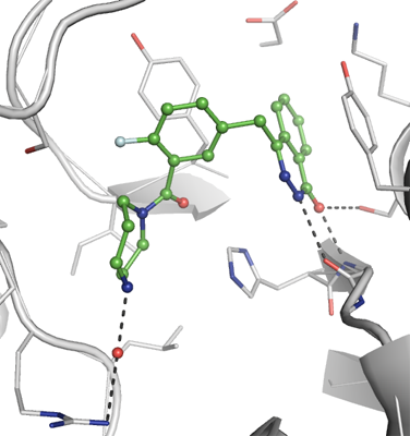
PARP3
PDB:3C49
Revision
Revision Type:created
Revised by:created
Revision Date:created
Entry Clone Accession:BC014260
Entry Clone Source:Mammalian Gene Collection
SGC Clone Accession:Tag:N-terminal hexahistidine tag with integrated TEV protease cleavage site: mhhhhhhssgvdlgtenlyfq*sm
Host:E.coli BL21(DE3) (Novagen)
Construct
Prelude:Sequence:mhhhhhhssgvdlgtenlyfq*smKRVQPCSLDPATQKLITNIFSKEMFKNTMALMDLDVKKMPLGKLSKQQIARGFEALEALEEALKGPTDGGQSLEELSSHFYTVIPHNFGHSQPPPINSPELLQAKKDMLLVLADIELAQALQAVSEQEKTVEEVPHPLDRDYQLLKCQLQLLDSGAPEYKVIQTYLEQTGSNHRCPTLQHIWKVNQEGEEDRFQAHSKLGNRKLLWHGTNMAVVAAILTSGLRIMPHSGGRVGKGIYFASENSKSAGYVIGMKCGAHHVGYMFLGEVALGREHHINTDNPSLKSPPPGFDSVIARGHTEPDPTQDTELELDGQQVVVPQGQPVPCPEFSSSTFSQSEYLIYQESQCRLRYLLEVH
Vector:pNIC-Bsa4
Growth
Medium:Antibiotics:Procedure:Cells from a glycerol stock were grown in 60 ml TB supplemented with 8 g/l glycerol, 100 µg/ml kanamycin and 34 µg/ml chloramphenicol at 37 ºC overnight. The overnight culture (60 ml) was used to inoculate 6 x 750 ml TB supplemented with 8 g/l glycerol and 50 µg/ml kanamycin. The cultures were grown in TunAir flasks at 37 ºC until OD600 reached ~1. The cultures were down-tempered to 18 ºC over a period of 1 hour before target expression was induced by addition of 0.5 mM IPTG. Expression was allowed to continue overnight and cells were harvested the following morning by centrifugation (5,500 x
g, 10 min, 4 ºC). The resulting cell pellet (150 g wet cell weight) was resuspended in lysis buffer (2 ml/g cell pellet), supplemented with three tablets of Complete EDTA-free protease inhibitor (Roche Applied Science). The cell suspension was stored at -80 ºC.
Purification
ProcedureColumnsIMAC 1: Ni-charged 5 ml HisTrap FF crude (GE Healthcare)
IMAC 2: Ni-charged 1 ml HiTrap chelating HP (GE Healthcare)
Gel filtration column: HiLoad 16/60 Superdex 75 Prep Grade (GE Healthcare)
Purification
Purification of the protein was performed as a two step process on an ÄKTApurifier system (GE Healthcare). Prior to purification, columns were equilibrated with IMAC wash1 buffer and gel filtration buffer, respectively. The filtered lysate was loaded onto the 5 ml HisTrap FF crude column and washed with IMAC wash1 buffer followed by IMAC wash2 buffer. Bound protein was eluted from the IMAC column with IMAC elution buffer and concentrated to 2.5 ml before loaded onto the gel filtration column. Fractions containing the target protein were pooled and fresh TCEP was added to a final concentration of 2 mM. The protein concentration was determined to 5.3 mg/ml in a volume of 13 ml.
Tag removal
The N-terminal histidine tag was proteolytically removed by incubating the target protein with His-tagged TEV protease at a molar ratio of protein:protease 30:1 at 4 ºC over the weekend. The proteolytic reaction went to completion, as judged by SDS-PAGE. The sample was diluted to 30 ml with IMAC wash2 buffer and thef imidazole concentration was adjusted to 25 mM. Target protein was purified from tag and protease by passing the reaction mixture over a 1 ml HiTrap chelating HP column (GE Healthcare) pre-equilibrated with IMAC wash2 buffer. The cleaved protein was collected in flow-through and concentrated to a volume of 10 ml. 5 ml of the cleaved protein was loaded onto the gel filtration column pre-equilibrated with gel filtration buffer. Fractions containing the target protein was concentrated to 40.2 mg/ml in 0.3 ml and stored at -80 ºC. The identity of the protein was confirmed by mass spectrometry.
Extraction
ProcedureThe cell suspension was quickly thawed in water. Cells were disrupted by sonication (Vibra-Cell, Sonics) at 80% amplitude for 3 min effective time (pulsed 4s on, 4s off). After sonication 6000 U Benzonase (Merck) was added to the sample and cell debris was removed by centrifugation (49,000 x
g, 30 min, 4 ºC). The supernatant was decanted and filtered through a 0.45 µm syringe filter.
Concentration:LigandMassSpec:Crystallization:Needle clusters of PARP3 were grown in a grid screen based on known conditions. Notably, seeding was not necessary. Crystals were obtained by the hanging drop vapour diffusion method in a 24-well plate containing 300 µl well solution. 2 µl of the protein solution (diluted to 13.5 mg/ml) was mixed with 1 µl of well solution consisting of 50 mM bis-Tris-propane pH 7.0 and 1.9 M DL-malic acid. The plate was incubated at 20 ºC and crystals appeared in 2 days. A single crystal was separated from the cluster and transferred to a drop containing 0.5 mM KU0058948 (stock 5 mM in DMSO), 25 mM bis-Tris-propane pH 7.0, 1.9 M DL-malic acid, and 0.25 M NaCl and incubated over night. The crystals were quickly transferred to cryo solution containing soaking solution (with 50 µM KU0058948) and 19 % glycerol. and flash frozen in liquid nitrogen.
NMR Spectroscopy:Data Collection:Data was collected at ESRF (ID14-2).
Data Processing:The structure was solved by molecular replacement using MOLREP with PARP3 structure (2PA9) as a search model. The inhibitor was easily located at the active site. The structure was refined with PHENIX and coordinates and structure factors were deposited in PDB with accession code 3C49.