TULP1
PDB:3C5N
Revision
Revision Type:created
Revised by:created
Revision Date:created
Entry Clone Accession:BC032714
Entry Clone Source:Mammalian Gene Collection
SGC Clone Accession:Tag:N-terminal hexahistidine tag with integrated TEV protease cleavage site: mhhhhhhssgvdlgtenlyfq*sm
Host:E.coli BL21(DE3) (Novagen)
Construct
Prelude:Sequence:mhhhhhhssgvdlgtenlyfq*smEPREFVLRPAPQGRTVRCRLTRDKKGMDRGMYPSYFLHLDTEKKVFLLAGRKRKRSKTANYLISIDPTNLSRGGENFIGKLRSNLLGNRFTVFDNGQNPQRGYSTNVASLRQELAAVIYETNVLGFRGPRRMTVIIPGMSAENERVPIRPRNASDGLLVRWQNKTLESLIELHNKPPVWNDDSGSYTLNFQGRVTQASVKNFQIVHADDPDYIVLQFGRVAEDAFTLDYRYPLCALQAFAIALSSFDG
Vector:pNIC-Bsa4
Growth
Medium:Antibiotics:Procedure:Cells from a glycerol stock were grown in 50 ml LB supplemented with 50 µg/ml kanamycin at 30 ºC overnight. The overnight culture (15 ml) was used to inoculate 2 x 750 ml TB supplemented with 8 g/l glycerol, 50 µg/ml kanamycin and approximately 50 µl BREOX FMT 30 anti-foam solution (Cognis Performance Chemicals UK Ltd). The cultures were grown in TunAir flasks at 37 ºC until OD600 reached ~1.1. The flasks were down-tempered to 18 ºC over a period of 1 hour before target expression was induced by addition of 0.5 mM IPTG. Expression was allowed to continue overnight and cells were harvested the following morning by centrifugation (4,400 x
g, 10 min, 4 ºC). The resulting cell pellet (38.3 g wet cell weight) was resuspended in lysis buffer (1 ml/g cell pellet), supplemented with two tablets of Complete EDTA-free protease inhibitor (Roche Applied Science) and a knife edge of lysozyme (Sigma). The cell suspension was stored at -80 ºC.
Purification
ProcedureColumns
IMAC: Ni-charged 1 ml HiTrap Chelating HP (GE Healthcare)
Gel filtration column: HiLoad 16/60 Superdex 75 Prep Grade (GE Healthcare)
Procedure
Purification of the protein was performed as a two step process on an ÄKTAprime system (GE Healthcare). Prior to purification, columns were equilibrated with IMAC wash1 buffer and gel filtration buffer, respectively. The filtered lysate was loaded onto the Ni-charged HiTrap Chelating column and washed with IMAC wash1 buffer followed by IMAC wash2 buffer. Bound protein was eluted from the IMAC column with IMAC elution buffer and loaded onto the gel filtration column. Fractions containing the target protein were pooled and the protein was subsequently concentrated in a Vivapore 5 centrifugal filter device, 7,500 NMWL (Sartorius) to 24.7 mg/ml in a volume of 0.4 ml. The identity of the protein was confirmed by mass spectrometry.
Extraction
ProcedureThe cell suspension was quickly thawed in water and 2000 U Benzonase was added. Cells were disrupted by high-pressure homogenization (TC5-0612W-332, Stansted fluid power LTD) and cell debris was removed by centrifugation (49,100 x
g, 20 min, 4 ºC). The supernatant was decanted and filtered through a 0.45 µm flask filter.
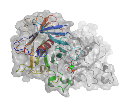
Concentration:LigandMassSpec:Crystallization:Crystals were obtained by the sitting drop vapour diffusion method in a 96-well plate. 1 µl protein solution (diluted to 20 mg/ml) including 1 mM myo-inositol 1,4,5 P3 was mixed with 0.5 µl well solution consisting of 10% (w/v) PEG 8000 and 10% (w/v) PEG 1000. The plate was incubated at 20 ºC and crystals appeared within 12 days. The crystals were quickly transferred to a cryo solution consisting of well solution complemented with 15% 2,3-butanediol and 5 mM myo-inositol 1,4,5 P3, and flash frozen in liquid nitrogen.
NMR Spectroscopy:Data Collection:Data was collected at BESSY beamline 14.2.
Data Processing:The data was integrated and scaled with XDS and XSCALE. The 1.8 Å structure was solved using 2FIM as the starting model in space group P21 and unit cell a=56.3, b=83.6, c=57.7, α9=0.00, β=94.01, γ=90.00. The model was refined using PHENIX and manually built using Coot. Residuals were Rfactor 0.189 and Rfree 0.221.