NUDT16
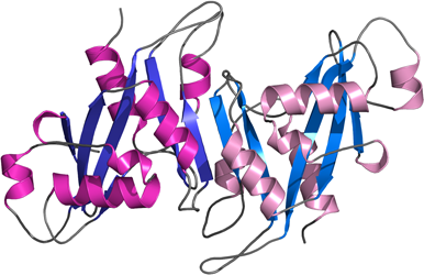
PDB:3COU
Revision
Revision Type:created
Revised by:created
Revision Date:created
Entry Clone Accession:BC031215
Entry Clone Source:Mammalian Gene Collection
SGC Clone Accession:NUDT16A-k001
Tag:N-terminal hexahistidine tag with integrated TEV protease cleavage site: mhhhhhhssgvdlgtenlyfq*sm
Host:E.coli BL21(DE3) R3 pRARE, where R3 denotes a derivative of BL21(DE3) resistant to a strain of T1 bacteriophage (SGC Oxford) and the pRARE plasmid originating from the Rosetta strain (Novagen) supplies tRNAs for rare codons.
Construct
Prelude:
Sequence:mhhhhhhssgvdlgtenlyfq*smAGARRLELGEALALGSGWRHVCHALLYAPDPGMLFGRIPLRYAILMQMRFDGRLGFPGGFVDTQDRSLEDGLNRELREELGEAAAAFRVERTDYRSSHVGSGPRVVAHFYAKRLTLEELLAVEAGATRAKDHGLEVLGLVRVPLYTLRDGVGGLPTFLENSFIGSAREQLLEALQDLGLLQSGSISGLKIPAHH
Vector:pNIC-Bsa4
Growth
Medium:
Antibiotics:
Procedure:Cells from a glycerol stock were grown in 10 ml TB supplemented with 8 g/l glycerol, 100 µg/ml kanamycin and 34 µg/ml chloramphenicol at 30 ºC overnight. 10 ml of the overnight culture were used to inoculate 0.75 l TB supplemented with 8 g/l glycerol, 50 µg/ml kanamycin and approximately 100 µl PPG P2,000 81380 anti-foam solution (Fluka). The culture was grown in a LEX bioreactor system (Harbinger Biotechnology) at 37 ºC until OD600 reached ~2. The bottle was down-tempered to 18 ºC over a period of 1 hour before target expression was induced by addition of 0.5 mM IPTG. Expression was allowed to continue overnight and cells were harvested the following morning by centrifugation (4,400 x g, 10 min, 4 ºC). The resulting cell pellet (17.7 g wet cell weight) was resuspended in lysis buffer (1.5 ml/g cell pellet), supplemented with 1000 U Benzonase (Merck) and 0.5 tablet of Complete EDTA-free protease inhibitor (Roche Applied Science). The cell suspension was stored at -80 ºC.
Purification
Procedure
Columns
IMAC: Ni-charged 1 ml HiTrap Chelating HP (GE Healthcare)
Gel filtration column: HiLoad 16/60 Superdex 75 Prep Grade (GE Healthcare)
Procedure
Purification of the protein was performed as a two step process on an ÄKTAxpress system (GE Healthcare). Prior to purification, columns were equilibrated with IMAC wash1 buffer and gel filtration buffer, respectively. The filtered lysate was loaded onto the HiTrap Chelating column and washed with IMAC wash1 buffer followed by IMAC wash2 buffer. Bound protein was eluted from the IMAC column with IMAC elution buffer and automatically loaded onto the gel filtration column. Fractions containing the target protein were pooled and fresh TCEP was added to a final concentration of 2 mM. The protein was subsequently concentrated using an Amicon Ultra-15 centrifugal filter device with 10,000 NMWL (Millipore) to 30.1 mg/ml in a volume of 0.75 ml. The identity of the protein was confirmed by mass spectrometry.
Extraction
Procedure
The cell suspension was quickly thawed in water and diluted to 68 ml with lysis buffer. Cells were disrupted by sonication (Vibra-Cell, Sonics) at 80% amplitude for 3 min effective time (pulsed 4s on, 4s off) and cell debris was removed by centrifugation (49,000 x g, 20 min, 4 ºC). The supernatant was decanted and filtered through a 0.45 µm flask filter.
Concentration:
Ligand
MassSpec:
Crystallization:Crystals were obtained by the sitting drop vapour diffusion method in a 96-well plate. 0.1 µl of the protein sample (30 mg/ml) was mixed with 0.1 µl of well solution consisting of 0.1 M CHES pH 9.5 and 20% (w/v) PEG 8000. The plate was incubated at 4 ºC and pyramidal crystals appeared within 7 days. The crystal was quickly transferred to cryo solution consisting of 0.1 M CHES pH 9.5, 21% (w/v) PEG 8000, 0.3 M NaCl and 20% glycerol, and flash-frozen in liquid nitrogen.
NMR Spectroscopy:
Data Collection:50.5º were collected by frame of 0.5º on the beamline BL14.1 at BESSY (0.9795Å).
Data Processing:The crystal belonged to space group F23 with cell parameters a=b=c=142.13 Å and α=β=γ= 90º. Data was processed and scaled using XDS and Scala respectively. The structure was solved by molecular replacement using PHASER. The probe was a model of NUDT16 provided by Swiss-Model based upon the structure of the nuclear snoRNA decapping protein X29 of Xenopus laevis (PDB-code 1U20). Data in the interval 19.7 Â 1.8 Å resolution was used for refinement. First steps of refinement were carried out by simulated annealing using CNS and automated model building starting from existing model using Arp/Warp. After these two steps, R and Rfree reached 19.5% and 24.5%.respectively. The final model was obtained by iterative cycles of manual building in COOT and refinement in REFMAC5. At the end of the refinement the values for R and Rfree were 18.4% and 21.7% respectively.