GLS
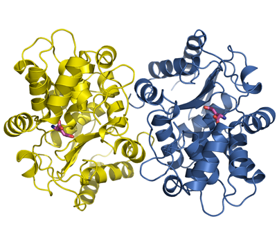
PDB:3CZD
Revision
Revision Type:created
Revised by:created
Revision Date:created
Entry Clone Accession:Entry Clone Source:In-house cloning
SGC Clone Accession:Tag:N-terminal hexahistidine tag with integrated TEV protease cleavage site: mhhhhhhssgvdlgtenlyfq*sm
Host: E.coli BL21(DE3) R3 pRARE, where R3 denotes a derivative of BL21(DE3) resistant to a strain of T1 bacteriophage (SGC Oxford) and the pRARE plasmid originating from the Rosetta strain (Novagen) supplies tRNAs for rare codons.
Construct
Prelude:Sequence:mhhhhhhssgvdlgtenlyfq*smIPDFMSFTSHIDELYESAKKQSGGKVADYIPQLAKFSPDLWGVSVCTVDGQRHSTGDTKVPFCLQSCVKPLKYAIAVNDLGTEYVHRYVGKEPSGLRFNKLFLNEDDKPHNPMVNAGAIVVTSLIKQGVNNAEKFDYVMQFLNKMAGNEYVGFSNATFQSERESGDRNFAIGYYLKEKKCFPEGTDMVGILDFYFQLCSIEVTCESASVMAATLANGGFCPITGERVLSPEAVRNTLSLMHSCGMYDFSGQFAFHVGLPAKSGVAGGILLVVPNVMGMMCWSPPLDKMGNSVKGIHFCHDLVSLCNFHNYDNL
Vector:pNIC-Bsa4
Growth
Medium:Antibiotics:Procedure:Freshly transformed cells were used to inoculate 6 x 10 ml TB supplemented with 8 g/l glycerol, 100 µg/ml kanamycin and 34 µg/ml chloramphenicol at 30 °C overnight. The overnight cultures were used to inoculate 6 x 0.75 l TB supplemented with 8 g/l glycerol, 50 µg/ml kanamycin and approximately 200 µl PPG P2,000 81380 anti-foam solution (Fluka). The cultures were grown in TunAir flasks at 37 °C until OD600 reached ~1.5. The flasks were down-tempered to 18 °C over a period of 1 hour before target expression was induced by addition of 0.5 mM IPTG. Expression was allowed to continue overnight and cells were harvested the following morning by centrifugation (4,400 x
g, 10 min, 4 °C). The resulting cell pellet (111.3 g wet cell weight) was resuspended in lysis buffer (1 ml/g cell pellet) supplemented with one tablet of Complete EDTA-free protease inhibitor (Roche Applied Science) and stored at -80 °C.
Purification
ProcedureColumns
IMAC: Ni-charged 5 ml HiTrap Chelating HP (GE Healthcare)
Gel filtration column: HiLoad 26/60 Superdex 200 Prep Grade (GE Healthcare)
Procedure
Purification of the protein was performed as a two step process on an ÄKTAxpress system (GE Healthcare). Prior to purification, columns were equilibrated with IMAC wash1 buffer and gel filtration buffer, respectively. The filtered lysate was loaded onto the Ni-charged HiTrap Chelating column and washed with IMAC wash1 buffer followed by IMAC wash2 buffer. Bound protein was eluted from the IMAC column with IMAC elution buffer and automatically loaded onto the gel filtration column. Fractions containing the target protein were pooled and subsequently concentrated in an Amicon Ultra-15 centrifugal filter device, 10,000 NMWL (Millipore) to 22.3 mg/ml in a volume of 1 ml.
Tag removal
The N-terminal histidine tag was proteolytically removed by incubating the target protein (22.3 mg/ml in 1.0 ml) with His-tagged TEV protease at a molar ratio of 30:1 at 4 ºC for 72 hours. The proteolytic reaction did not reach completion, as judged by SDS-PAGE, why additionally TEV protease (20:1 final molar ratio) was added to the protein:protease mix and incubated at 20 ºC overnight. The target protein was subsequently purified from tag and protease by passing it over a Ni-charged 1 ml HisTrap FF column (GE Healthcare) pre-equilibrated with IMAC wash1 buffer. The cleaved protein was concentrated and the buffer was changed to GF buffer in an Amicon Ultra-15 centrifugal filter device, 10,000 NMWL (Millipore). The final protein concentration was determined to 19.8 mg/ml in a volume of 0.9 ml. The identity of the protein was confirmed by mass spectrometry.
Extraction
ProcedureThe cell suspension was quickly thawed in water and supplemented with 4000 U Benzonase (Merck). Cells were disrupted by sonication (Vibra-Cell, Sonics) at 80% amplitude for 3 min effective time (pulsed 4s on, 4s off) and cell debris was removed by centrifugation (49,000 x
g, 25 min, 4 ºC). The supernatant was decanted and filtered through a 0.45 µm flask filter.
Concentration:LigandMassSpec:Crystallization:Crystals were obtained by the hanging drop vapour diffusion method in a 24-well plate containing 500 µl well solution. 1.4 µl of the protein solution (19.8 mg/ml) including 10 mM L-glutamate was mixed with 0.7 µl of well solution consisting of 0.1 M bis-Tris-propane pH 7.0 and 1.4 M lithium sulfate. The plate was incubated at 20 ºC and small crystals appeared after 2 days. After growing for one week the crystal was quickly transferred to cryo solution containing well solution, 0.2 M NaCl and 25% glycerol, and flash frozen in liquid nitrogen.
NMR Spectroscopy:Data Collection:Diffraction data to 2.4 Å resolution was collected at MAX-lab beamline I911-3.
Data Processing:The structure was solved by molecular replacement using the structure of glutaminase from
Micrococcus luteus K-3 (PDB entry 2DFW) as search model. The space group was I4122 with cell dimensions a=b=139.52 Å c=153.95 Å. One monomer was located in the asymmetric unit. Refmac5 was used for refinement and Coot for model building. Refinement using TLS-parameters were done in the later stages. Data in the interval 29.0-2.40 Å resolution was used and at the end of the refinement the values for R=18.85% and Rfree=21.00%. Coordinates for the crystal structure were deposited in the Protein Data Bank, accession code 3CZD.