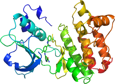
EPHA7
PDB:3DKO
Revision
Revision Type:created
Revised by:created
Revision Date:created
Entry Clone Accession:NP_004431
Entry Clone Source:epha7.001.998.CDV.pOT50
SGC Clone Accession:epha7.590.899.125E12 (SDC125E12)
Tag:N-terminal tag: M
C-terminal tag: AHHHHHH
Host:BL21 (DE3)
Construct
Prelude:
Sequence:MQEGDEELYFHFKFPGTKTYIDPETYEDPNRAVHQFAKELDASCIKIERVIGAGEFGEVCSGRLKLPGKRDVAVAIKTLKVGYTEKQRRDFLCEASIMGQFDHPNVVHLEGVVTRGKPVMIVIEFMENGALDAFLRKHDGQFTVIQLVGMLRGIAAGMRYLADMGYVHRDLAARNILVNSNLVCKVSDFGLSRVIEDDPEAVYTTTGGKIPVRWTAPEAIQYRKFTSASDVWSYGIVMWEVMSYGERPYWDMSNQDVIKAIEEGYRLPAPMDCPAGLHQLMLDCWQKERAERPKFEQIVGILDKMIRNPNSAHHHHHH
Vector:pNIC-CH
Growth
Medium:TB
Antibiotics:
Procedure:A 250 ml flask containing LB (Sigma L7658) supplemented with 50 ug/ ml kanamycin (BioShop Canada KAN 201) was inoculated from a glycerol stock of the bacteria. The flask was shaken overnight (16 hours) at 250 rpm at 37 degC.
Using the Lex system, a 2L bottle (VWR 89000-242) containing 1800 ml of TB (Sigma T0918) supplemented with 1.5% glycerol, 50 ug/ ml kanamycin and 600 ul antifoam 204 (Sigma A-8311) was inoculated with 50 ml overnight LB culture, and incubated at 37 degC. The temperature of the media was reduced to 15 degC one hour prior to induction and induced at OD600 = 6 with 100 uM isopropyl-thio-β-D-galactopyranoside (BioShop Canada IPT 001). Cultures were aerated overnight (16 hours) at 15 degC, and cell pellets collected by centrifugation and frozen at -80 degC.
Purification
Procedure
Unclarified lysate was mixed with 2-3 mL of HisLink Protein Purification Resin (Promega V8821) per 40 mL lysate. The mixture was incubated with mixing for at least 20 minutes at 4 degC. The lysate was spun at 500 xg for 5 minutes, and the supernatant was decanted. The remaining resin was washed with 45 mL of cold wash buffer, allowing 5 minutes to settle between washes, until the supernatant was clear (usually 3-5 washes). The washed resin was transferred to a gravity column and further washed with 1 column volumes (approx. 5 mL) of wash buffer at approximately 3 mL/min. Samples were eluted from the resin by exposure to 2-3 column volumes (approx. 10 mL) of elution buffer.
An XK 16x65 column (part numbers 18-1031-47 and 18-6488-01, GE Healthcare) packed with HighLoad Superdex 200 resin (10-1043-04, GE Healthcare) was pre-equilibrated with gel filtration buffer for 1.5 column volumes using an AKTAxpress (18-6645-05, GE Healthcare) at a flow rate of 3 mL/min. The eluate sample from the IMAC step (approx. 10 mL) was loaded onto the column at 1.5 mL/min, and 2mL fractions were collected into 96-well plates (VWR 40002-012) using peak fractionation protocols). Fractions observed by a UV absorption chromatogram to contain the protein were pooled.
Ligand
Name: ALW-II-49-7
Chemical Formula: C21H17F3N4O2
Exact Mass: 414.13
Chemical name: 5-(2-methyl-5-(3-(trifluoromethyl)phenylcarbamoyl)phenylamino)nicotinamide
Extraction
Procedure
Frozen cell pellet contained in bags (Beckman 369256) obtained from 2L of culture were thawed by soaking in warm water. Each cell pellet was resuspended in 25-40 mL lysis buffer and homogenized using an Ultra-Turrax T8 homogenizer (IKA Works) at maximal setting for 30-60 seconds per pellet. Cell lysis was accomplished by sonication (Virtis408912, Virsonic) on ice: the sonication protocol was 10 sec pulse at half-maximal frequency (5.0), 10 second rest, for 10 minutes total sonication time per pellet.
Concentration:Purified proteins were concentrated using 15 mL concentrators with a 5,000 molecular weight cut-off (Amicon Ultra-15, UFC900524, Millipore) at 3750 rpm, typically resulting in a final concentration of 10-15 mg/mL.
Ligand
MassSpec:
Crystallization:Co-Crystallization with inhibitors
The pooled sample from gel-filtration was concentrated to 40 micromolar, supplemented with DMSO (5% final concentration), and mixed at 4 degC, dropwise, with inhibitor (stock concentration at 800 micromolar inhibitor, 5% DMSO, in Gel Filtration Buffer) to reach 80 micromolar final inhibitor concentration. After a 2 hour, 4 degC, followed by 30 minute 22 degC incubation, the sample was spun (to remove precipitates) and the supernatant was concentrated to 15 mg/ml.
Crystallizationconditions
Crystals of the EphA7 kinase domain were grown at 298 K using the sitting drop method by mixing equal volumes of 25% PEG3350, 0.1 M NH4SO4, 0.1 M HEPES pH 7.5 and 15 mg/mL protein sample. The crystals were cryoprotected by transferring the crystals to a drop containing mother liquor to which MPD was added to 25%.
NMR Spectroscopy:
Data Collection:
Data Processing: