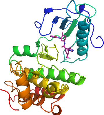
EPHA3
PDB:3DZQ
Revision
Revision Type:created
Revised by:created
Revision Date:created
Entry Clone Accession:BC063282
Entry Clone Source:MGC
SGC Clone Accession:epha3.606.947.033G08 (SDC033G08)
Tag:N-terminal tag: MGSSHHHHHHSSGLVPRGS
Host:BL21 (DE3)
Construct
Prelude:
Sequence:mgsshhhhhhssglvprgsTQTVHEFAKELDATNISIDKVVGAGEFGEVCSGRLKLPSKKEISVAIKTLKVGYTEKQRRDFLGEASIMGQFDHPNIIRLEGVVTKSKPVMIVTEYMENGSLDSFLRKHDAQFTVIQLVGMLRGIASGMKYLSDMGYVHRDLAARNILINSNLVCKVSDFGLSRVLEDDPEAAYTTRGGKIPIRWTSPEAIAYRKFTSASDVWSYGIVLWEVMSYGERPYWEMSNQDVIKAVDEGYRLPPPMDCPAALYQLMLDCWQKDRNNRPKFEQIVSILDKLIRNPGSLKIITSAAARPSNLLLDQSNVDITTFRTTGDWLNGVWTAHCKEIFTGVEYSSCDTIAKIS
Vector:pET28aLIC
Growth
Medium:
Antibiotics:
Procedure:Competent BL21 (DE3) cells (Invitrogen, C6000-03) were transformed and grown using the LEX system (HarbingerBiotech) at 37 °C in 2L bottles (VWR, 89000-242) containing 1800 ml of TB (Sigma, T0918) supplemented with 150 mM glycerol, 100 µM Kanamycin and 600 µl antifoam 204 (Sigma A-8311). When the OD(600) reached a value of about 6.0, the temperature was reduced to 15 °C, and one hour later the culture was induced with 200 µM IPTG (BioShop, IPT001) and incubated overnight (16 hours) at 15 °C. Cell pellets were collected by centrifugation (12,227 xg, 20 mins), frozen in liquid nitrogen, and stored at -80 °C.
Purification
Procedure
The remaining resin was washed with 45 mL of cold wash buffer, allowing 5 minutes to settle between washes, until the supernatant was clear (usually 3-5 washes). The washed resin was transferred to a gravity column and further washed with 1 column volumes (approx. 5 mL) of wash buffer at approximately 3 mL/min. Samples were eluted from the resin by exposure to 2-3 column volumes (approx. 10 mL) of elution buffer. An XK 16x65 column (part numbers 18-1031-47 and 18-6488-01, GE Healthcare) packed with HighLoad Superdex 200 resin (10-1043-04, GE Healthcare) was pre-equilibrated with gel filtration buffer for 1.5 column volumes using an AKTAxpress (18-6645-05, GE Healthcare) at a flow rate of 3 mL/min. The eluate sample from the IMAC step (approx. 10 mL) was loaded onto the column at 1.5 mL/min, and 2mL fractions were collected into 96-well plates (VWR 40002-012) using peak fractionation protocols). Fractions observed by a UV absorption chromatogram to contain the protein were pooled. Purified proteins were concentrated using 15 mL concentrators with a 5,000 molecular weight cut-off (Amicon Ultra-15, UFC900524, Millipore) at 3750 rpm, typically resulting in a final concentration of 10-15 mg/mL. Mass-spectroscopy by LCMS showed that this protein undergoes post-translational modifications such as degradation, phosphorylation, and mercaptoethanoylation.
Extraction
Procedure
Frozen cell pellet contained in bags (Beckman 369256) obtained from 2L of culture were thawed by soaking in warm water. Each cell pellet was resuspended in 25-40 mL lysis buffer and homogenized using an Ultra-Turrax T8 homogenizer (IKA Works) at maximal setting for 30-60 seconds per pellet. Cell lysis was accomplished by sonication (Virtis408912, Virsonic) on ice: the sonication protocol was 10 sec pulse at half-maximal frequency (5.0), 10 second rest, for 10 minutes total sonication time per pellet. Unclarified lysate was mixed with 2-3 mL of HisLink Protein Purification Resin (Promega V8821) per 40 mL lysate. The mixture was incubated with mixing for at least 20 minutes at 4 degC. The lysate was spun at 500 xg for 5 minutes, and the supernatant was decanted.
Concentration:The pooled sample from gel-filtration was concentrated to 50 mM, supplemented with DMSO (5% final concentration), and mixed at 4 degC, dropwise, with ALW-II-38-3 (C23H18F3N5O3, 469.14 g/mol, N-(2-methyl-5-(3-(4-methyl-1H-imidazol-1-yl)-5-(trifluoromethyl)-benzamido)-phenyl)-isoxazole-5-carboxamide, stock concentration at 800 mM inhibitor, 5 % DMSO, in Gel Filtration Buffer) to reach 80 mM final inhibitor concentration. After a 2 hour, 4 degree, followed by 30 minute 22 degC incubation, the sample was spun (to remove precipitates) and the supernatant was concentrated to 20 mg/ml.
Ligand
MassSpec:
Crystallization:Crystals of the EphA3 kinase domain were grown at 298 K using the sitting drop method by mixing equal volumes of 30% PEG3350, 0.2 M NH4SO4, 0.1 M Na CaCo pH 6.5 and 20 mg/mL protein sample. The crystals were cryoprotected by transferring the crystals to a drop containing mother liquor to which glycerol was added to 20%.
NMR Spectroscopy:
Data Collection:Diffraction data from a crystal of EphA3 kinase domain with the inhibitor ALW-II-38-3 was collected using a FR-E home-source X-ray generator. All data sets were integrated and scaled using either the HKL2000 or HKL3000 program packages. The co-crystal structure of EphA3 in complex with inhibitor ALW-II-38-3 was solved by difference Fourier techniques using the previously determined apo EphA3 kinase domain structure (PDB entry 2GSF), and refined as in the previous inhibitor structure. Parameters for Translation/liberation/screw (TLS) refinement were generated using the TLSMD web server.
Data Processing:The coordinates and structure factors were deposited on 2008.07.30 into the RCSB PDB database with ID code 3DZQ with the following authors: John R. Walker, Farisa Syeda, Nathanael Gray, W. Mansoor, Johan Weigelt, Chas Bountra, Aled M. Edwards, Cheryl H. Arrowsmith, Alexey Bochkarev, and Sirano Dhe-Paganon.