FLJ10324
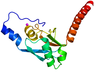
PDB:3EC8
Revision
Revision Type:created
Revised by:created
Revision Date:created
Entry Clone Accession:BC117317
Entry Clone Source:Mammalian Gene Collection
SGC Clone Accession:FLJ10324A-k012
Tag:N-terminal hexahistidine tag with integrated TEV protease cleavage site: mhhhhhhssgvdlgtenlyfq*sm
Host:E.coli BL21(DE3) R3 pRARE, where R3 denotes a derivative of BL21(DE3) resistant to a strain of T1 bacteriophage (SGC Oxford) and the pRARE plasmid originating from the Rosetta strain (Novagen) supplies tRNAs for rare codons.
Construct
Prelude:Sequence:mhhhhhhssgvdlgtenlyfq*smDPAELSTQLSAPGVLKVFGDSVCTGTHYKSVLATGTSSARELVKEALERYALDPRQAGQYVLCDVVGQAGDAGQRWQARCFRVFGDSEKPLLIQELWKPREGLSRRFELRKRSDVEELAAKEVDTITAGINAQARRLQRSRAK
Vector:pNIC-Bsa4
Growth
Medium:Antibiotics:Procedure:Native proteinCells from a glycerol stock were grown in 20 ml TB supplemented with 8 g/l glycerol, 100 µg/ml kanamycin and 34 μg/ml chloramphenicol at 37 °C overnight. The overnight culture was then used to inoculate 1.5 l TB supplemented with 8 g/l glycerol, 50 µg/ml kanamycin, 34 μg/ml chloramphenicol and approximately 200 µl Dow Corning anti-foam RD emulsion 63213 4D (BDH Silicone Products). The culture was grown in a LEX bioreactor system (Harbinger Biotechnology) at 37 °C until OD600 reached ~2. The bottle was down-tempered to 18 °C over a period of 1 hour before expression of target protein was induced by addition of 0.5 mM IPTG. Expression was allowed to continue overnight and cells were harvested the following morning by centrifugation (5,500 x
g, 10 min, 4 °C). The resulting cell pellet (21.6 g wet cell weight) was resuspended in lysis buffer (3 ml/g cell pellet) supplemented with 2000 U Benzonase (Merck) and one tablet of Complete EDTA-free protease inhibitor (Roche Applied Science) and stored at -80 °C.
SeMet labeled protein
Cells from glycerol stock were grown in 100 ml SOB supplemented with 50 µg/ml kanamycin and 34 μg/ml chloramphenicol at 30 °C overnight. 90 ml of the overnight culture was used to inoculate 12 bottles with 750 ml minimal medium (without amino acids) supplemented with 50 µg/ml kanamycin and approximately 200 µl Dow Corning anti-foam RD emulsion 63213 4D (BDH Silicone Products). The culture was grown at 37 °C until OD600 reached ~0.6. The bottles were down-tempered to 18 °C during 30 min before amino acids were added. After additionally 30 min, expression of target protein was induced by addition of 0.5 mM IPTG. The expression was allowed to continue at 18 °C overnight. Cells were harvested the following morning by centrifugation (4,400 x g, 20 min, 4 °C). The resulting cell pellet (30 g from 9 liter culture) was resuspended in lysis buffer (2 ml/g cell pellet) supplemented with 1500 U Benzonase (Merck) and one tablet of Complete EDTA-free protease inhibitor (Roche Applied Science) and stored at -80 °C.
Composition of Minimal media: 25 mM Na2HPO4, 25 mM KH2PO4, 50 mM NH4Cl, 5 mM Na2SO4, 0.4% (w/v) glucose, 2mM MgSO4, 0.1mM CaCl2, 0.5 µM MnCl2, 10 µM FeSO4.
Mix of amino acids added (per liter): 100 mg each of lysine, threonine, phenylalanine and 50 mg each of leucine, isoleucine, valine, L(+)-selenomethionine.
Purification
ProcedureColumnsIMAC: Ni-charged 1 ml HiTrap Chelating HP (GE Healthcare)
Gel filtration column: HiLoad 16/60 Superdex 75 Prep Grade (GE Healthcare)
Procedure
Purification of the native protein was performed as a two step process on an ÄKTAxpress system (GE Healthcare). Prior to purification, columns were equilibrated with IMAC wash1 buffer and gel filtration buffer, respectively. The filtered lysate was loaded onto the Ni-charged HiTrap Chelating column and washed with IMAC wash1 buffer followed by IMAC wash2 buffer. Bound protein was eluted from the IMAC column with IMAC elution buffer and automatically loaded onto the gel filtration column. Fractions containing the target protein were pooled and subsequently concentrated using an Amicon Ultra-15 centrifugal filter device, 5,000 NMWL (Millipore) to 14.8 mg/ml in a volume of 1.5 ml and stored at -80 ºC. Purification of the SeMet enriched protein was performed in basically the same way. The filtered lysate was purified by IMAC and gel filtration chromatography followed by a concentration step. The final SeMet-protein concentration was 39.3 mg/ml in a volume of 1.2 ml. The identities of the proteins were confirmed by mass spectrometry.
Extraction
ProcedureThe cell suspension was quickly thawed in water. Cells were disrupted by sonication (VibraCell, Sonics) at 80% amplitude for 3 min effective time (puls 4s on, 4s off) and cell debris was removed by centrifugation (49,000 x
g, 20 min, 4 ºC). The supernatant was decanted and filtered through a 0.45 µm flask filter.
Concentration:LigandMassSpec:Crystallization:The crystals were obtained by the sitting drop vapour diffusion method in a 96-well plate. 0.1 µl of the protein solution (14.8 mg/ml) was mixed with 0.1 µl of well solution consisting of 0.2 M ammonium dihydrogen phosphate, 0.1 M Tris pH 8.5 and 50% MPD. The plates were incubated at 4 ºC. The derivative crystals were obtained by soaking crystals in Pb and Hg salts for several minutes. SeMet crystals were produced in the same way by mixing 0.2 µl of the SeMet-protein solution (diluted with GF buffer to 15 mg/ml) with 0.1 µl of well solution consisting of 0.4 M ammonium dihydrogen phosphate, 0.1 M Tris pH 8.5 and 35% (v/v) MPD. The plate was incubated at 4 ºC and crystals were obtained after 28 days. The crystals were quickly transferred to cryo solution containing well solution, 0.3 M NaCl and 20% glycerol and flash frozen in liquid nitrogen.
NMR Spectroscopy:Data Collection:Data sets were collected at BESSY (BL14-2)
Data Processing:The crystals belonged to space group P3112 with the cell parameters 46.5 46.5 135.2 90 90 120. The structure was solved by MIRAS autoSHARP giving three data files (native-2.8 Å, Hg-2.8 Å and Pb-2.4 Å) that had been scaled together in XDS & XSCALE. Heavy atom files came from shelx(cd). The native dataset was initially collected as SeMet data and was re-indexed in XDS-CORRECT prior to scaling it to the other two in XSCALE. Refmac5 was used for refinement and Coot for model building. Refinement using TLS-parameters was done at a later stage. Data in the interval 19.7 - 2.6 Å resolution was used, and after refinement the R values were R = 22.7% and Rfree = 27%. Coordinates were deposited in the Protein Data Bank, accession code 3EC8.