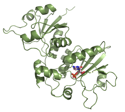
PCYT2
PDB:3ELB
Revision
Revision Type:created
Revised by:created
Revision Date:created
Entry Clone Accession:BC000351
Entry Clone Source:Mammalian Gene Collection
SGC Clone Accession:Tag:N-terminal hexahistidine tag with integrated TEV protease cleavage site: mhhhhhhssgvdlgtenlyfq*sm
Host:E.coli BL21(DE3) R3 pRARE, where R3 denotes a derivative of BL21(DE3) resistant to a strain of T1 bacteriophage (SGC Oxford) and the pRARE plasmid originating from the Rosetta strain (Novagen) supplies tRNAs for rare codons.
Construct
Prelude:Sequence:mhhhhhhssgvdlgtenlyfq*smGGRRAVRVWCDGCYDMVHYGHSNQLRQARAMGDYLIVGVHTDEEIAKHKGPPVFTQEERYKMVQAIKWVDEVVPAAPYVTTLETLDKYNCDFCVHGNDITLTVDGRDTYEEVKQAGRYRECKRTQGVSTTDLVGRMLLVTKAHHSSQEMSSEYREYADSFGKCPGGRNPWTGVSQFLQTSQKIIQFASGKEPQPGETVIYVAGAFDLFHIGHVDFLEKVHRLAERPYIIAGLHFDQEVNHYKGKNYPIMNLHERTLSVLACRYVSEVVIGAPYAVTAELLSHFKVDLVCHGKTEIIPDRDGSDPYQEPKRRGIFRQIDSGSNLTTDLIVQRIITNRLEY
Vector:pNIC-Bsa4
Growth
Medium:Antibiotics:Procedure:Cells from glycerol stock were grown in 2 x 40 ml LB supplemented with 100 µg/ml kanamycin, and 34 μg/ml chloramphenicol at 30 °C overnight. The overnight cultures (60 ml) were used to inoculate 6 x 1.5 l minimal medium (without amino acids) supplemented with 50 µg/ml kanamycin, 34 μg/ml chloramphenicol and approximately 200 µl PPG P2,000 81380 anti-foam solution (Fluka). The cultures were grown in a LEX bioreactor system (Harbinger Biotechnology) at 37 °C until OD
600 of ~0.6 when the bottles were down tempered to a 18 °C. After 10 minutes, amino acids (Van Duyne, G. D.,
J. Mol. Biol. 229, 105-124 (1993)) were added and after additionally one hour, expression of target protein was induced by addition of 0.5 mM IPTG. Expression was allowed to continue overnight and cells were harvested the following morning by centrifugation (4400 x
g, 15 min, 4 °C). The resulting cell pellet (43.1 g wet cell weight) was resuspended in lysis buffer (2 ml/g cell pellet), supplemented with 1.5 tablet of Complete EDTA-free protease inhibitor (Roche Applied Science). The cell suspension was stored at -80 °C. Selenomethionine enriched protein was grown in minimal medium containing: 25 mM Na
2HPO
4, 25 mM KH
2PO
4, 50 mM NH4Cl, 5 mM Na
2SO4, 0.4% (w/v) glucose, 2 mM MgSO4, 0.1 mM CaCl2, 1.0 µM MnCl2, 10 µM FeSO4, pH ~7. Mix of amino acids added (per liter culture): 100 mg each of lysine, threonine, phenylalanine and 50 mg each of leucine, isoleucine, valine, L(+)-selenomethionine.
Purification
ProcedureColumns
IMAC: Ni-charged 1 ml HiTrap Chelating HP (GE Healthcare)
Gel filtration column: HiLoad 16/60 Superdex 200 Prep Grade
Procedure
Purification of the protein was performed as a two step process on an ÄKTAxpress system (GE Healthcare). Prior to purification, columns were equilibrated with IMAC wash1 buffer and gel filtration buffer, respectively. The filtered lysate was loaded onto two HiTrap Chelating columns connected in series and washed with IMAC wash1 buffer followed by IMAC wash2 buffer. Bound protein was eluted from the IMAC columns with IMAC elution buffer and automatically loaded onto the gel filtration column. Fractions containing the target protein were pooled and the protein was concentrated using a Vivaspin 20 centrifugal filter device with 10,000 MWCO (Sartorius) to 5.1 mg/ml in a volume of 1.1 ml.
Tag removal
The N-terminal histidine tag was proteolytically removed by incubating the target protein with His-tagged TEV protease (van den Berg, S., J. Biotech 121, 291-298 (2006)) at a molar ratio of 30:1 at 20 ºC overnight. The protein sample was diluted with IMAC wash1 buffer without TCEP, to lower the TCEP concentration in the sample. The target protein was purified from tag and protease by passing the reaction mixture over a Ni-charged 1 ml HisTrap HP column (GE Healthcare) pre-equilibrated with IMAC wash1 buffer. The cleaved protein was concentrated and the buffer was changed to GF buffer using a Vivaspin 20 centrifugal filter device with 30,000 MWCO (Sartorius). The final protein concentration was determined to 13.0 mg/ml in a volume of 0.13 ml. The identity of the protein was confirmed by mass spectrometry and selenomethionine was fully incorporated at all 7 methionine positions in the labeled protein.
Extraction
ProcedureThe cell suspension was quickly thawed in water and 3000 U Benzonase (Merck) was added. Cells were disrupted by sonication (Vibra-Cell, Sonics) at 80% amplitude for 3 min effective time (pulsed 4s on, 4s off) and cell debris was removed by centrifugation (49,000 x
g, 20 min, 4 ºC). The supernatant was decanted and filtered through a 0.45 µm flask filter.
Concentration:LigandMassSpec:Crystallization:SeMet labeled crystals were obtained by the sitting drop vapour diffusion method in a 96-well plate. 0.2 µl of the protein solution (diluted to 12.2 mg/ml) including 10 mM CTP and 10 mM MgCl2 was mixed with 0.1 µl of well solution consisting of 0.2 M magnesium formate pH 7.5, 20% PEG 3350. The plate was incubated at 20 ºC and crystals appeared within 2 days. Cryo solution containing well solution, 0.2 M NaCl and 20% glycerol was added directly to the drop. Crystals were mounted and flash-frozen in liquid nitrogen.
NMR Spectroscopy:Data Collection:Data sets were collected at BESSY (BL14-1)
Data Processing:Diffraction data to 2.0 Å resolution were collected. The structure was solved by single anomalous diffraction (SAD) using the incorporated selenomethionines. SHELXC/D/E software was used to locate and refine the sites. The space group was P41212 with cell dimensions a = b = 74.18 Å and c = 139.72 Å. One monomer was located in the asymmetric unit. ARP/wARP was initially used for automatic model building; Refmac5 was used for refinement and Coot for manual model building. Data in the interval 19.7 - 2.0 Å was used, and after refinement the R values were R = 19.8% and Rfree = 24.2%. Coordinates were deposited in the Protein Data Bank with accession code 3ELB.