SKIL
PDB:3EQ5
Revision
Revision Type:created
Revised by:created
Revision Date:created
Entry Clone Accession:BC059386
Entry Clone Source:Mammalian Gene Collection
SGC Clone Accession:Tag:N-terminal hexahistidine tag with integrated TEV protease cleavage site: mhhhhhhssgvdlgtenlyfq*sm
Host: E.coli BL21(DE3) R3 pRARE, where R3 denotes a derivative of BL21(DE3) resistant to a strain of T1 bacteriophage (SGC Oxford) and the pRARE plasmid originating from the Rosetta strain (Novagen) supplies tRNAs for rare codons.
Construct
Prelude:Sequence:mhhhhhhssgvdlgtenlyfq*smPSDSSTELTQTVLEGESISCFQVGGEKRLCLPQVLNSVLREFTLQQINTVCDELYIYCSRCTSDQLHILKVLGILPFNAPSCGLITLTDAQRLCNALLRPRT
Vector:pNIC-BSA4
Growth
Medium:Antibiotics:Procedure:Cells from a glycerol stock were grown in 10 ml TB supplemented with 8 g/l glycerol, 100 µg/ml kanamycin and 34 µg/ml chloramphenicol at 30 °C overnight. The overnight culture (10 ml) was used to inoculate 0.75 l TB supplemented with 8 g/l glycerol, 50 µg/ml kanamycin and approximately 100 µl PPG P2,000 81380 anti-foam solution (Fluka). The culture was grown in a LEX bioreactor system (Harbinger Biotechnology) at 37 °C until OD600 reached ~2. The bottle was down-tempered to 18 °C over a period of 1 hour before target expression was induced by addition of 0.5 mM IPTG. Expression was allowed to continue overnight and cells were harvested the following morning by centrifugation (4,400 x
g, 10 min, 4 °C). The resulting cell pellet (9.0 g wet cell weight) was resuspended in lysis buffer (3 ml/g cell pellet), supplemented with 1000 U Benzonase (Merck) and 0.5 tablet of Complete EDTA-free protease inhibitor (Roche Applied Science). The cell suspension was stored at -80 °C.
Purification
ProcedureColumnsIMAC: Ni-charged 1 ml HiTrap Chelating HP (GE Healthcare)
Gel filtration column: HiLoad 16/60 Superdex 75 Prep Grade (GE Healthcare)
Procedure
Purification of the protein was performed as a two step process on an ÄKTAxpress system (GE Healthcare). Prior to purification, columns were equilibrated with IMAC wash1 buffer and gel filtration buffer, respectively. The filtered lysate was loaded onto the Ni-charged HiTrap Chelating column and washed with IMAC wash1 buffer followed by IMAC wash2 buffer. Bound protein was eluted from the IMAC column with IMAC elution buffer and automatically loaded onto the gel filtration column. Fractions containing the target protein were pooled and fresh TCEP was added to a final concentration of 2 mM. The protein was subsequently concentrated using a Amicon Ultra-15 centrifugal filter device with 5,000 NMWL (Millipore) to 42.7 mg/ml in a volume of 1.2 ml.
Tag removal
The N-terminal histidine tag was proteolytically removed by incubating the target protein with His-tagged TEV protease (van den Berg, S., J. Biotech 121, 291-298 (2006)) at a molar ratio of 30:1 at 4 ºC overnight. The proteolytic reaction went to completion, as judged by SDS-PAGE. Target protein was purified from tag and protease by passing the reaction mixture over a Ni-charged 1 ml HiTrap Chelating HP column (GE Healthcare) pre-equilibrated with IMAC wash1 buffer. The cleaved protein was concentrated and the buffer was changed to GF buffer using a centrifugal filter device with 5,000 NMWL. The final protein concentration was determined to 19.6 mg/ml in a volume of 0.7 ml. The identity of the protein was confirmed by mass spectrometry.
Extraction
ProcedureThe cell suspension was quickly thawed in water and diluted to 68 ml with lysis buffer. Cells were disrupted by sonication (Vibra-Cell, Sonics) at 80% amplitude for 3 min effective time (pulsed 4s on, 4s off) and cell debris was removed by centrifugation (49,000 x
g, 20 min, 4 ºC). The supernatant was decanted and filtered through a 0.45 µm flask filter.
Concentration:LigandMassSpec:Crystallization:Crystals were obtained by the sitting drop vapour diffusion method in a 96-well plate. 0.1 µl of the protein solution (19.6 mg/ml) was mixed with 0.2 µl of well solution consisting of 0.2 M ammonium nitrate and 20% (w/v) PEG 3350. The plate was incubated at 4 ºC and rhombohedral crystals appeared after 15 to 29 days. After 73 days, the crystal was transferred to cryo solution consisting of 20 mM HEPES, 300 mM NaCl, 20% glycerol, 0.2 M ammonium nitrate and 20% (w/v) PEG 3350, pH 7.5, and flash-frozen in liquid nitrogen.
NMR Spectroscopy:Data Collection:125° was collected by frame of 0.5° at ESRF (ID29) (0.97623 Å).
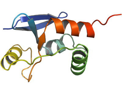
Data Processing:The crystal belonged to space group C2 with the cell parameters a=210.820, b=70.120, c=116.280, α=γ=90.00 and β=100.95. Data was processed and scaled using XDS and XSCALE. PHASER found 7 correct monomers in the asymmetric unit using the structure of the Dachshund-homology domain of human SKI (PDB-code: 1SBX) as a probe. This solution was refined in CNS using simulated annealing. The resulting model was used as a probe in a new query in PHASER and resulted in the discovery of two more monomers. Analysis of the crystallographic contacts allowed to build 3 additional monomers in the density map with Coot, resulting in the final asymmetric unit of 12 monomers. Data in the interval 19.95 - 2.45 Å resolution was used for refinement. Iterative cycles of manual building in both Coot and O and restrained refinement in REFMAC5 were performed. Final TLS refinement using 3 TLS groups for 11 monomer and 4 for monomer E led to the values of 23.2% and 27.3% for R and Rfree respectively.