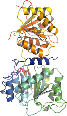
DDX19
PDB:3EWS
Revision
Revision Type:created
Revised by:created
Revision Date:created
Entry Clone Accession:BC003626
Entry Clone Source:Mammalian Gene Collection
SGC Clone Accession:Tag:N-terminal hexahistidine tag with integrated TEV protease cleavage site: mhhhhhhssgvdlgtenlyfq*sm
Host:E.coli BL21(DE3) R3 pRARE, where R3 denotes a derivative of BL21(DE3) resistant to a strain of T1 bacteriophage (SGC Oxford) and the pRARE plasmid originating from the Rosetta strain (Novagen) supplies tRNAs for rare codons.
Construct
Prelude:Sequence:mhhhhhhssgvdlgtenlyfq*smEDRAAQSLLNKLIRSNLVDNTNQVEVLQRDPNSPLYSVKSFEELRLKPQLLQGVYAMGFNRPSKIQENALPLMLAEPPQNLIAQSQSGTGKTAAFVLAMLSQVEPANKYPQCLCLSPTYELALQTGKVIEQMGKFYPELKLAYAVRGNKLERGQKISEQIVIGTPGTVLDWCSKLKFIDPKKIKVFVLDEADVMIATQGHQDQSIRIQRMLPRNCQMLLFSATFEDSVWKFAQKVVPDPNVIKLKREEETLDTIKQYYVLCSSRDEKFQALCNLYGAITIAQAMIFCHTRKTASWLAAELSKEGHQVALLSGEMMVEQRAAVIERFREGKEKVLVTTNVCARGIDVEQVSVVINFDLPVDKDGNPDNETYLHRIGRTGRFGKRGLAVNMVDSKHSMNILNRIQEHFNKKIERLDTDDLDEIE
Vector:pNIC-Bsa4
Growth
Medium:Antibiotics:Procedure:Cells from a glycerol stock were grown in 30 ml TB supplemented with 8 g/l glycerol, 100 µg/ml kanamycin and 34 µg/ml chloramphenicol at 30 °C overnight. The overnight culture (10 ml) was used to inoculate 750 ml TB supplemented with 8 g/l glycerol, 50 µg/ml kanamycin and approximately 100 µl PPG P2,000 81380 anti-foam solution (Fluka). The culture was grown in a LEX bioreactor system (Harbinger Biotechnology) at 37 °C until OD600 reached ~2.2. The bottle was down-tempered to 18 °C over a period of 1 hour before target expression was induced by addition of 0.5 mM IPTG. Expression was allowed to continue overnight and cells were harvested the following morning by centrifugation (4,500 x
g, 10 min, 4 °C). The resulting cell pellet (8.4 g wet cell weight) was resuspended in lysis buffer (3.0 ml/g cell pellet), supplemented with 1000 U Benzonase (Merck) and 0.5 tablet of Complete EDTA-free protease inhibitor (Roche Applied Science). The cell suspension was stored at -80 °C.
Purification
ProcedureColumnsIMAC: Ni-charged 1 ml HiTrap Chelating HP (GE Healthcare)
Gel filtration column: HiLoad 16/60 Superdex 200 Prep Grade (GE Healthcare)
Procedure
Purification of the protein was performed as a two step process on an ÄKTAxpress system (GE Healthcare). Prior to purification, columns were equilibrated with IMAC wash1 buffer and gel filtration buffer, respectively. The filtered lysate was loaded onto the Ni-charged HiTrap Chelating column and washed with IMAC wash1 buffer followed by IMAC wash2 buffer. Bound protein was eluted from the IMAC column with IMAC elution buffer and automatically loaded onto the gel filtration column. Fractions containing the target protein were pooled and fresh TCEP was added to a final concentration of 2 mM. The protein was subsequently concentrated using an Amicon Ultra-15 centrifugal filter device with 30,000 NMWL (Millipore) to 20.6 mg/ml in a volume of 1 ml. The identity of the protein was confirmed by mass spectrometry.
Extraction
ProcedureThe cell suspension was briefly thawed in water. Cells were disrupted by sonication (Vibra-Cell, Sonics) at 80% amplitude for 3 min effective time (pulsed 4s on, 4s off) and cell debris was removed by centrifugation (49,000 x
g, 20 min, 4 ºC). The supernatant was decanted and filtered through a 0.45 µm flask filter.
Concentration:LigandMassSpec:Mass spectra of DDX19 showed a sample of correct mass.
Crystallization:Crystals were obtained by the sitting drop vapour diffusion method in a 96-well plate. Protein solution, ADP and MgCl2 were mixed to yield a final sample composition of 18.3 mg/ml protein, 20 mM ADP and 10 mM MgCl2. 0.1 µl of the protein sample was then mixed with 0.1 µl of well solution consisting of 0.1 M bis-Tris, pH 5.5, 0.1 M ammonium acetate, 17% PEG-10000. The plate was incubated at 4 ºC and crystals appeared within 8 days. After additionally one week, cryo solution containing well solution and 20% glycerol was added directly to the drop. Crystals were mounted and flash-frozen in liquid nitrogen.
NMR Spectroscopy:Data Collection:Data was collected at BESSY (beam line BL14-2) to 2.7 Å resolution.
Data Processing:Data was processed with XDS. The crystal belonged to space group p21 with a=81.6 Å b=47.6 Å c=124.7 Å and β=95.4°. The structure was solved by PHASER using 1FUU as a search model. Domains were searched for separately and they were edited according to the sequence alignment with Chainsaw before molecular replacement. The structure was refined with PHENIX. TLS parameters were refined using individual domains as rigid groups. Model building was done using Coot. The structure finally refined to R-factors of 0.19 and 0.26 for R and Rfree, respectively. Coordinates were deposited in the Protein Data Bank with an accession code 3EWS.