PARP3
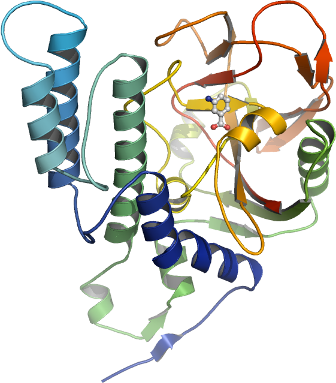
PDB:3FHB
Revision
Revision Type:created
Revised by:created
Revision Date:created
Entry Clone Accession:Entry Clone Source:Mammalian Gene Collection
SGC Clone Accession:Tag:N-terminal hexahistidine tag with integrated TEV protease cleavage site: mhhhhhhssgvdlgtenlyfq*sm
Host:E.coli BL21(DE3) (Novagen)
Construct
Prelude:Sequence:mhhhhhhssgvdlgtenlyfq*smKRVQPCSLDPATQKLITNIFSKEMFKNTMALMDLDVKKMPLGKLSKQQIARGFEALEALEEALKGPTDGGQSLEELSSHFYTVIPHNFGHSQPPPINSPELLQAKKDMLLVLADIELAQALQAVSEQEKTVEEVPHPLDRDYQLLKCQLQLLDSGAPEYKVIQTYLEQTGSNHRCPTLQHIWKVNQEGEEDRFQAHSKLGNRKLLWHGTNMAVVAAILTSGLRIMPHSGGRVGKGIYFASENSKSAGYVIGMKCGAHHVGYMFLGEVALGREHHINTDNPSLKSPPPGFDSVIARGHTEPDPTQDTELELDGQQVVVPQGQPVPCPEFSSSTFSQSEYLIYQESQCRLRYLLEVH
Vector:pNIC-Bsa4
Growth
Medium:Antibiotics:Procedure:Cells from a glycerol stock were plated on LB-agar with appropriate antibiotics. 5-10 colonies were used to inoculate 40 ml TB supplemented with 8 g/l glycerol and 50 µg/ml kanamycin and grown at 37 ºC overnight. The overnight culture (20 ml) was used to inoculate 1.5 l TB supplemented with 8 g/l glycerol and 50 µg/ml kanamycin and 200 µl BREOX FMT 30 anti-foam solution (Cognis Performance Chemicals UK Ltd). The culture was grown in a LEX bioreactor system (Harbinger Biotechnology) at 37 ºC until OD600 reached ~1.2. The culture was down-tempered to 18 ºC over a period of 1 hour before target expression was induced by addition of 0.5 mM IPTG. Expression was allowed to continue overnight and cells were harvested the following morning by centrifugation (5,500 x
g, 10 min, 4 ºC). The resulting cell pellet (30 g wet cell weight) was resuspended in lysis buffer (1 ml/g cell pellet), supplemented with 0.5 tablet of Complete EDTA-free protease inhibitor (Roche Applied Science). The cell suspension was stored at -80 ºC.
Purification
ProcedureColumns
IMAC: Ni-charged 1 ml HiTrap chelating HP (GE Healthcare)
Gel filtration column: HiLoad 16/60 Superdex 200 Prep Grade (GE Healthcare)
Procedure
Purification of the protein was performed as a two step process on an ÄKTAxpress system (GE Healthcare). Prior to purification, columns were equilibrated with IMAC wash1 buffer and gel filtration buffer, respectively. The filtered lysate was loaded onto the Ni-charged HiTrap Chelating column and washed with IMAC wash1 buffer followed by IMAC wash2 buffer. Bound protein was eluted from the IMAC column with IMAC elution buffer and automatically loaded onto the gel filtration column. Fractions containing the target protein were pooled and fresh TCEP was added to a final concentration of 2 mM. The protein concentration was determined to 1.25 mg/ml in a volume of 9.5 ml.
Tag removal
The N-terminal histidine tag was proteolytically removed by incubating the target protein with His-tagged TEV protease in a molar ratio of protein:protease 50:1 at 4 ºC for 72 hours. The proteolytic reaction did not reach completion, as judged by SDS-PAGE. Target protein was purified from tag, protease and uncleaved protein by passing the reaction mixture over a 1 ml HisTrap FF crude column (GE Healthcare) pre-equilibrated with 20 mM HEPES, 500 mM NaCl, 10% Glycerol, 10 mM Imidazole, 2 mM TCEP, pH 7.5. The cleaved protein was collected in flow-through. The buffer was changed to GF-buffer with (2mM TCEP), concentrated to 19.4 mg/ml in 0.23 ml and stored at -80 ºC. The identity of the protein was confirmed by mass spectrometry.
Extraction
ProcedureThe cell suspension was quickly thawed in water and 1500 U Benzonase (Merck) was added to the sample. Cells were disrupted by sonication (Vibra-Cell, Sonics) at 80% amplitude for 3 min effective time (pulsed 4s on, 4s off) and cell debris was removed by centrifugation (49,000 x
g, 20 min, 4 ºC). The supernatant was decanted and filtered through a 0.45 µm flask filter.
Concentration:LigandMassSpec:Crystallization:Needle-like crystals of PARP3 were found during initial screening with the JCSG+ crystal screen. However, well diffracting and useful crystals could only be obtained using seeding techniques. Crystals were obtained by the hanging drop vapour diffusion method in a 24-well plate containing 500 µl well solution. 1.5 µl of the protein solution (19 mg/ml) containing 10 mM 3-amino benzoic acid was mixed with 1.5 µl of well solution consisting of 0.1 M bis-Tris-propane pH 7.0 and 1.8 M DL-malic acid. After one day incubation the drops were streak-seeded from the initial crystal hits. The plate was incubated at 20 ºC and crystals appeared in 3 days. Cryo solution containing well solution and 30% glycerol was added to the drop and crystals were flash frozen in liquid nitrogen.
NMR Spectroscopy:Data Collection:Diffraction data was collected at beamline BM14 at the ESRF synchrotron radiation facility in Grenoble, France. Data was indexed and integrated in space group P21 with the XDS package. The cell parameters are a=55.24 Å, b=56.70 Å, c=56.48 Å, b=112.86°.
Data Processing:The structure was solved by molecular replacement using PHASER with an ensemble generated by MrBUMP and consisting of human and mouse structures of PARP1 as search model. The asymmetric unit contained one protein monomer. Refmac5 was used for refinement and Coot for model building. In the refinement, data in the interval 19.92-2.30 Å resolution was used and the progress of refinement was followed by decreasing R and Rfree values. At the end of the refinement the R values were: R= 0.1876 and Rfree= 0.2566. A Ramachandran plot showed 97.7% of all residues in favored regions and no outliers in the disallowed regions. Coordinates for the crystal structure were deposited in the Protein Data Bank, accession code 3FHB.