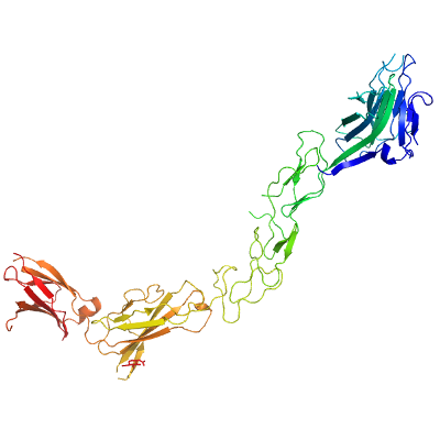
EPHA2
PDB:3FL7
Revision
Revision Type:created
Revised by:created
Revision Date:created
Entry Clone Accession:BC037166
Entry Clone Source:MGC
SGC Clone Accession:epha2.023.531.121A10 (SDC121A10)
Tag:N-terminal tag: AAPEHHHHHHDYDIPTTENLYFQGAMDAA
Host:
Construct
Prelude:
Sequence:aapehhhhhhdydipttenlyfqgamdaaQGKEVVLLDFAAAGGELGWLTHPYGKGWDLMQNIMNDMPIYMYSVCNVMSGDQDNWLRTNWVYRGEAERIFIELKFTVRDCNSFPGGASSCKETFNLYYAESDLDYGTNFQKRLFTKIDTIAPDEITVSSDFEARHVKLNVEERSVGPLTRKGFYLAFQDIGACVALLSVRVYYKKCPELLQGLAHFPETIAGSDAPSLATVAGTCVDHAVVPPGGEEPRMHCAVDGEWLVPIGQCLCQAGYEKVEDACQACSPGFFKFEASESPCLECPEHTLPSPEGATSCECEEGFFRAPQDPASMPCTRPPSAPHYLTAVGMGAKVELRWTPPQDSGGREDIVYSVTCEQCWPESGECGPCEASVRYSEPPHGLTRTSVTVSDLEPHMNYTFTVEARNGVSGLVTSRSFRTASVSINQTEPPKVRLEGRSTTSLSVSWSIPPPQQSRVWKYEVTYRKKGDSNSYNVRRTEGFSVTLDDLAPDTTYLVQVQALTQEGQGAGSKVHEFQTLSPEG
Vector:pFHMSP-LIC-N
Growth
Medium:
Antibiotics:
Procedure:The plasmid epha2.023.531.121A10 (SDC121A10) was transformed into DH10Bac E. coli cells (Invitrogen, 10361-012), and a mini-prep was performed to obtain recombinant viral DNA bacmid. SF9 cells (Invitrogen, 11496-015) were transfected with bacmid using Cellfectin reagent (Invitrogen, 10362-010) and recombinant baculovirus was generated. Viral stock was amplified from P1 to P3.
Sf9 cells were grown in Serum Free Medium (HyClone SFX-Insect, SH3027802) at density of 3.5 million cells per milliliter of media and with viability not less then 97 % were infected with 10 mL of P3 viral stock for each 1 L of cell culture. Cell culture medium was collected after 3-4 days of incubation on a shaker at 100 rpm and 27 ºC when cells viability dropped to 45-65 %.
Purification
Procedure
A 3.2 L volume of medium was mixed with 30 ml pre-equilibrated NiNTA Superflow beads (Qiagen, 30450) and stirred (Talboys, 134-2) on ice for 1 hour. The resin was transferred to a 100 ml gravity column, washed with 300 ml of Wash Buffer and the protein was eluted with 30-40 ml of Elution Buffer. A second round of NiNTA batch absorption may have been performed for increased yield. The eluate was dialyzed against 50-100 X volume of buffer 50 mM Tris pH8.0 and 0.15 M NaCL.
Extraction
Procedure
The cultured medium was centrifuged at 14,000 xg for 15 minutes, and the pH of the supernatant was adjusted to 7.5 at room temperature by adding Titrating Buffer. PMSF (Bioshop, PMS 123.50) and benzamidine (Sigma, B6506-100G) were added to final concentrations of 1 and 2 mM, respectively.
Concentration:Purified protein was concentrated using 15 mL concentrators with an appropriate molecular weight cut-off (Amicon Ultra-15 10,000 MWCO, Millipore) to a final value of 10 mg/mL. Average yield was about 2 mg/L. Coomassie-stained SDS-PAGE showed that the product was pure and mass-spectroscopy by LC/MS (Agilent 1100 Series) showed that the protein was glycosylated.
Ligand
MassSpec:
Crystallization:Crystals were grown at 18 °C using the hanging drop method in greased VDX Plates (Hampton Research, HR3-306) by mixing equal volumes of protein (8.6 mg/ml) and Crystallization Buffer (3 % PEG 4 K, 0.1 M NaAc, 0.1 M CaCo pH 5.5 with 0.5 M NDSB 256). Prior to dunking and storage in liquid nitrogen, suitable crystals were immersed in 1 uL CB and 1 uL cryoprotectant (20% (w/v) sucrose, 4% (w/v) glucose, 18% (v/v) glycerol and 18% (v/v) ethylene glycol).
NMR Spectroscopy:
Data Collection:The coordinates and structure factors have been deposited into the RCSB PDB database with ID code 3FL7 with the following PDB authors: John R. Walker, Laila Yermekbayeva, Alma Seitova, Christine Butler-Cole, Johan Weigelt, Chas Bountra, Aled M. Edwards, Cheryl H. Arrowsmith, Alexey Bochkarev, and Sirano Dhe-Paganon.
Data Processing: