NUDT6
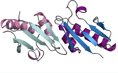
PDB:3FXT
Revision
Revision Type:created
Revised by:created
Revision Date:created
Entry Clone Accession:BC009842
Entry Clone Source:Mammalian Gene Collection
SGC Clone Accession:
Tag:N-terminal hexahistidine tag with integrated TEV protease cleavage site: mhhhhhhssgvdlgtenlyfq*sm
Host:E.coli BL21(DE3) R3 pRARE, where R3 denotes a derivative of BL21(DE3) resistant to a strain of T1 bacteriophage (SGC Oxford) and the pRARE plasmid originating from the Rosetta strain (Novagen) supplies tRNAs for rare codons.
Construct
Prelude:
Sequence:mhhhhhhssgvdlgtenlyfq*smDLQGELDRFGGISVRLARLDALDRLDAAAFQKGLQAAVQQWRSEGRTAVWLHIPILQSRFIAPAASLGFCFHHAESDSSTLTLWLREGPS
Vector:pNIC-Bsa4
Growth
Medium:
Antibiotics:
Procedure:Cells from a glycerol stock were grown in 15 ml TB supplemented with 8 g/l glycerol, 100 µg/ml kanamycin and 34 µg/ml chloramphenicol at 30 °C overnight. The overnight culture (15 ml) was used to inoculate 750 ml TB supplemented with 8 g/l glycerol, 50 µg/ml kanamycin and approximately 200 µl 204 Antifoam A6426 (Sigma). The culture was grown in a LEX bioreactor system (Harbinger Biotechnology) at 37 °C until OD600 reached ~2. The bottle was down-tempered to 18 °C over a period of 1 hour before target expression was induced by addition of 0.5 mM IPTG. Expression was allowed to continue overnight and cells were harvested the following morning by centrifugation (4,400 x g, 10 min, 4 °C). The resulting cell pellet (13.9 g wet cell weight) was resuspended in lysis buffer (2 ml/g cell pellet), supplemented with 1000 U Benzonase (Merck) and 0.5 tablet of Complete EDTA-free protease inhibitor (Roche Applied Science). The cell suspension was stored at -80 °C.
Purification
Procedure
Columns
IMAC: Ni-charged 1 ml HiTrap Chelating HP (GE Healthcare)
Gel filtration column: HiLoad 16/60 Superdex 75 Prep Grade (GE Healthcare)
Procedure
Purification of the protein was performed as a two step process on an ÄKTAxpress system (GE Healthcare). Prior to purification, columns were equilibrated with IMAC wash1 buffer and gel filtration buffer, respectively. The filtered lysate was loaded onto the Ni-charged HiTrap Chelating column and washed with IMAC wash1 buffer followed by IMAC wash2 buffer. Bound protein was eluted from the IMAC column with IMAC elution buffer and automatically loaded onto the gel filtration column. Fractions containing the target protein were pooled and fresh TCEP was added to a final concentration of 2 mM. The protein was subsequently concentrated using an Amicon Ultra-15 centrifugal filter device, 5,000 NMWL (Millipore) to 8 mg/ml in a volume of 1.2 ml. The identity of the protein was confirmed by mass spectrometry.
Extraction
Procedure
The cell suspension was quickly thawed in water and diluted with lysis buffer to a final volume of 68 ml. Cells were disrupted by sonication (Vibra-Cell, Sonics) at 80% amplitude for 3 min effective time (pulsed 4s on, 4s off) and cell debris was removed by centrifugation (49,100 x g, 20 min, 4 ºC). The supernatant was decanted and filtered through a 0.45 µm flask filter.
Concentration:
Ligand
MassSpec:
Crystallization:Native crystals were obtained by the hanging drop vapour diffusion method in a 24-well plate. Prior to setting up the plate, protein solution was mixed with chymotrypsin (Sigma-Aldrich) in a protease:protein molar ratio of 1:100. 1 µl of the chymotrypsin treated protein solution (8 mg/ml) was mixed with 1 µl of well solution consisting of 0.1 M Tris-HCl pH 7.5, 0.25 M trimethylamine and 19% PEG 2000 MME. The plate was incubated at 4 ºC and rhombohedral crystals appeared in two days. After one month, the crystals were briefly transferred to a cryo solution consisting of well solution complemented with 30% glycerol and flash frozen in liquid nitrogen.
Hg-derivatized crystals were obtained in the same way as described above except that the well solution contained 0.3 M trimethylamine. Protein was treated with chymotrypsin as previously described. After 20 days, crystals were transferred to a drop containing 0.1 M Tris-HCl pH 7, 0.3 M trimethylamine, 10% PEG 2000 MME, 1 mM HgAc and equilibrated against a well solution, containing the same solution as the drop but without HgAc, for 17 hours. The Hg-derivatized crystals were then briefly transferred to a cryo solution composed of 0.1 M Tris-HCl pH 7, 0.3 M trimethylamine, 10% PEG 2000 MME, 1 mM HgAc and 30% glycerol and flash frozen in liquid nitrogen.
Sm-derivatized crystals were obtained by the hanging drop vapour diffusion method in a 24-well plate. Prior to setting up crystallization trials, chymotrypsin was added to the protein as previously described. 2 µl of protein solution (8 mg/ml) was mixed with 1 µl of the well solution consisting of 0.1 M Tris-HCl pH 7.5, 14% PEG 2000 MME and incubated at 4 ºC. After 8 days, crystals were transferred to a drop containing 0.1 M Tris-HCl pH 7, 0.3 M trimethylamine, 10% PEG 2000 MME and 1 mM SmCl3 and equilibrated against a well solution, containing the same solution as in the drop but without SmCl3, for two weeks. Crystals were then briefly transferred to a cryo solution composed of 0.1 M Tris-HCl pH 7, 0.3 M trimethylamine, 10% PEG 2000 MME, 1 mM SmCl3 and 30% glycerol and flash frozen in liquid nitrogen.
NMR Spectroscopy:
Data Collection:Native dataset: 120° were collected by frame of 0.5° on the beamline ID29 at ESRF (0.97931 Å). Hg-derivatized dataset: 360° were collected by frame of 0.5° on the beamline ID29 at ESRF (1.0056 Å). Sm-derivatized dataset: 282.5° were collected by frame of 0.5° on the beamline BL14-2 at BESSY (1.8449 Å).
Data Processing:The three crystals used for structure determination appeared to be isomorphous and belonged to space group P64 2 2. The cell parameters for the native dataset were a=b=185.8 Å, c=129.2 Å, α=β=90.00° and γ=120°. Data was processed and scaled using XDS and XScale respectively. AutoSharp was able to find the correct MIRAS solution while restricted the resolution to native, Hg and Sm dataset to 2.3, 3.4 and 3 Å respectively. 11 Hg sites and 11 Sm sites were located. After solvent flattening and density modification, ArpWarp built and docked 575 residues belonging to 7 molecules in the asymetric unit leading to R and Rfree values of 0.24 and 0.33 respectively. An eighth monomer was placed in Coot and after adding water and restrained refinement cycles in Refmac, R and Rfree dropped to 0.24 and 0.29. The model was improved using Phenix (R/Rfree = 0.24/0.28). Final steps of refinment were processed by iterative cycles of manual building in Coot and restrained refinement in Refmac. Finally, TLS refinement led to a model whose R and Rfree values are 0.2 and 0.24 respectively (2 groups of TLS per monomer) and which was deposited in the PDB with accession id: 3FXT.