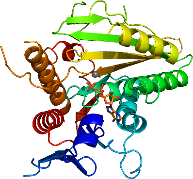
KIF13B
PDB:3GBJ
Revision
Revision Type:created
Revised by:created
Revision Date:created
Entry Clone Accession:SGC:25-G1
Entry Clone Source:CDV synthesized cDNA template
SGC Clone Accession:HPC092-C02
Tag:C-terminal His6
Host:BL21-DE3-V2R pRARE2
Construct
Prelude:KIF13B:S4-K351:cH6
N-terminal Methionine was cleavaged.
Sequence:SKVKVAVRIRPMNRRETDLHTKCVVDVDANKVILNPVNTNLSKGDARGQPKVFAYDHCFWSMDESVKEKYAGQDIVFKCLGENILQNAFDGYNACIFAYGQTGSGKSYTMMGTADQPGLIPRLCSGLFERTQKEENEEQSFKVEVSYMEIYNEKVRDLLDPKGSRQTLKVREHSVLGPYVDGLSKLAVTSYKDIESLMSEGNKSRTVAATNMNEESSRSHAVFKITLTHTLYDVKSGTSGEKVGKLSLVDLAGSERATKTGAAGDRLKEGSNINKSLTTLGLVISALADQSAGKNKNKFVPYRDSVLTWLLKDSLGGNSKTAMVATVSPAADNYDETLSTLRYADRAKhhhhhh
Vector:pET28a-LIC-CHis (GI:145307000), His6-tag was encoded in the primer used for PCR amplification. Vector digested with NcoI/BseRI, cloning used Clontech Infusion method.
Growth
Medium:Terrific Broth
Antibiotics:Kanamycin 50 µg/mL Chloramphenicol 25 µg/mL
Procedure:LEX Bubbling. The target protein was expressed in E. coli by inoculating 100 mL of overnight culture grown in Luria-Bertani medium into per 2L Terrific Broth medium in the presence of 50 µg/mL kanamycin and 50 µg/mL chloramphenicol at 37 °C. When OD600 reached 4.2, the temperature of the medium was lowered to 18 degC and the culture was induced with 1 mM IPTG. The cells were allowed to grow overnight before they were harvested and flash frozen in liquid nitrogen and stored at -80 °C.
Purification
Procedure
The lysate was centrigued at 15,000 rpm for 45 minutes and the supernatants were mixed with 4 mL 50% Ni-NTA beads, and incubated at 4 degC for 1 hours. The supernatant was then passed through a gravity column (Poly-Prep, Bio-Rad, catalog #731-1550) and the beads were washed using 50 mL binding buffer twice. The protein bound to beads were eluted using elution buffer twice in a total 30 mL volume. The flow-through was collected and loaded onto Supderdex-200 26/60 gel filtration column. Eluted fractions were pooled and then loaded onto MonoQ 10/50 GL prepacked column and eluted gradiently. Eluted fractions came out from around 23.2% Buffer B, were pooled and concentrated using an Amicon centrifugal filter (m.w. cut-off 10,000 ). MgCL2 was added to a final concentration of 4mM and ADP added to final 10 times molarity of protein concentration. The purity of the proteins was higher than 98% judged by SDS-PAGE.
Extraction
Procedure
Total frozen cells from 6L culture were thawed and resuspended in extraction buffer at 5mL buffer per 1g wet cells, and added 1mM PMSF/Benzomidine, 5U/ml of Benzonase (Sigma Catalog # E1014, 250U/µL), 0.5% CHAPS freshly, and supplemented with protease inhibitor cocktail (SIGMA Catalog # P8849), and lysed using sonication (100W 5min with 10s on and 10s off duty cycle).
Concentration:18.1 mg/mL
Ligand
Mg2+, ADPMassSpec:HPC092-C02: 39050.85, expected 39181.31, the N-terminal Methione was processed and cleavaged by E.coli.
Crystallization:Buffer for protein is 20mM HEPES pH7.0, 230 mM NaCl. Concentrated protein was added with MgCl2 to a final concentration of 4mM and 10 times molarity of ADP (vs. protein concentration) and setup for crystallization. Crystal used for data collection was grown in 17.7% PEG3350, 0.4M (NH4)2Citrate using 0.5 uL protein plus 0.5 uL well solution, sitting drop setting up. Crystal usually appear in 1-2 days. There are two crystal forms seen in the initial screen, diamond shaped or orthogonal rod. Crystal shape changed from rod-shaped to dimond for 18.6%PEG3350, 0.4M (NH4)2Citrate. Dimond shaped crystal never diffracts better than rod shaped crystals.
Cryoprotectant: 23%P3350 0.2M (NH4)2Citrate 0.3M NaCl 8%EG
NMR Spectroscopy:
Data Collection:
Data Processing: