Entry Clone Source: Ordered-synthetic |
Entry Clone Accession: n/a |
SGC Construct ID: TXNRD3A-c136 |
GenBank GI number: gi|51492609 |
Vector: pNIC-CTHF. Details [ PDF ]; Sequence [ FASTA ] or [ GenBank ]
|
Amplified construct sequence:
CTTAAGAAGGAGATATACTATGGCGCGCG
AAGAACTGCGTCGTCATCTGGTTGGACTG
ATTGAGCGCTCCCGTGTGGTAATCTTTTC
AAAGTCTTATTGTCCGCATAGTACGCGTG
TTAAAGAACTGTTTTCCAGCCTGGGTGTA
GAATGCAATGTGCTGGAGCTGGACCAGGT
GGATGATGGTGCCCGCGTTCAGGAAGTCC
TGTCTGAAATTACCAACCAGAAAACGGTC
CCGAACATTTTCGTGAATAAAGTGCACGT
CGGTGGCTGCGATCAGACTTTTCAGGCAT
ATCAGAGTGGGCTGCTGCAGAAACTGCTG
CAAGAAGACCTGGCCTATGATGCAGAGAA
CCTCTACTTCCAATCGCACCATCATCACC
ACCATGATTACAAGGATGACGACGATAAG
TGAGGATCC |
Tags and additions: TEV-cleavable (*), C-terminal histag. Tag sequence: aenlyfq*shhhhhhdykddddk |
Protein sequence (after TEV cleavage; tag sequence in lowercase):
MAREELRRHLVGLIERSRVVIFSKSYCPH
STRVKELFSSLGVECNVLELDQVDDGARV
QEVLSEITNQKTVPNIFVNKVHVGGCDQT
FQAYQSGLLQKLLQEDLAYDaenlyfq |
Host: ORIGAMI2 |
Growth medium, induction protocol: Medium: TB, 50 µg/ml Kanamycin and 10 µg/ml tetracycline. 2x2 liter TB in 5-L flasks were inoculated with 3x20 ml overnight culture and grown at 37°C until OD600 reached 2.5 . The temperature was then decreased to 18°C and the protein expression induced with 0.1 mM IPTG over night . The cells were collected by centrifugation and frozen at -80°C. |
Extraction buffer, extraction method: Lysis buffer: 50 mM HEPES pH 7.5, 500 mM NaCl, 10 mM Imidazole, 5% glycerol, 0.5 mM TCEP, 1 mM PMSF and 3 U/ml of Benzonase. Cell pellets were resuspended in a total volume of 250 ml lysis buffer. The cells were disrupted by high pressure (15 kpsi) and nucleic acids and cell debris removed by adding 0.15% PEI, followed by centrifugation for 60 minutes at 40000xg. The supernatant was further clarified by filtration (0.20 µm). |
Column 1: Ni-Sepharose FF, 3 ml (GE/Amersham Biosciences ) |
Buffers: Binding buffer: 50 mM HEPES pH 7.5, 500 mM NaCl, 10 mM Imidazole, 5% glycerol , 0.5 mM TCEP; Wash and elution buffers: 50 mM HEPES pH 7.5, 500 mM NaCl, 5% glycerol 10-250 mM imidazole. |
Procedure: The cell lysate was applied onto a 3 ml Ni-Sepharose FF column equilibrated with binding buffer. The column was subsequently washed with 30 ml of binding buffer and the protein eluted using a stepwise gradient of imidazole. All fractions were collected and analysed by SDS-PAGE. |
Column 2: Hiload 16/60 Superdex 200 prep grade 120 ml (GE/Amersham Biosciences). |
Buffers: 10 mM HEPES, pH 7.5, 500 mM NaCl, 5% glycerol, 0.5 mM TCEP. |
Procedure: The fractions eluted from the Ni-affinity chromatography were incubated with 0.5 mM TCEP and concentrated in amicon (3K) to 5 ml. The protein was filtered through a 0.20 µm PVDF filter and applied to a Superdex S200 column at 1 ml/min. The eluted proteins were collected in 1.8 ml fractions and analysed by SDS-PAGE. |
Enzymatic treatment: TEV cleavage. |
Column 3: Ni-Sepharose FF (TEV clean up) |
Buffer: 10 mM HEPES, pH 7.5, 500 mM NaCl, 5% glycerol |
Procedure: The Histidine-tag was cleaved with 150 µg of TEV protease per 10 mg protein at 4°C for 16 hours. The TEV cleaved protein was applied to a 0.5 ml Ni-sepharose column and the flow through collected. The column was washed with 5 ml buffer and the flow through and the wash fraction were analysed by SDS-PAGE. |
Concentration: The protein was concentrated in Amicon (3 K) to 12.3 mg/ml. The protein concentration was determined spectrophotometrically using the predicted molar extinction coefficient 5960 (M-1 cm-1). |
Mass spectrometry characterization: Two peaks of 13017.0 and 12886.1 Da were determined by ES-TOF for TXNRD3Ap005 which correspond respectively to the expected mass of the construct (13016.8 Da) and the protein lacking the N-terminal methionine (12885.8) |
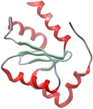
Crystallisation: The GLRX domain in TXNRD3A was crystallised by vapor diffusion at 20ºC in a sitting drop consisting of 100 nL protein (12.3 mg/ml) and 50 nL well solution. The drop was equilibrated against well solution containing 0.1 M Hepes pH 7.5, 2 M (NH4)2SO4. The crystal was transferred to a cryoprotectant composed of 20% ethylene glycol before flash-cooling in liquid nitrogen. |
Data Collection: Resolution: 2.2 Å; X-ray source: Synchrontron SLS-X10. |
Data Processing: Data were processed and scaled using MOSFLM and SCALA. Initial structure solution was obtained by molecular replacement using the program PHASER and the coordinate of glutaredoxin C1 of from populus tremula x tremuloides (pdb id: 2e7p) as a search model. Phases were improved by applying NCS averaging and solvent flattening using the program Parrot. Automated model building was performed using the program Buccaneer. The model was refined using REFMAC5 with manual rebuilding in COOT. The final model was refined against 2.21 Å with the final Rfact of 0.189 and Rfree of 0.233. |