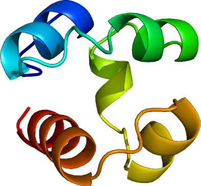
EPHA1
PDB:3HIL
Revision
Revision Type:created
Revised by:created
Revision Date:created
Entry Clone Accession:NP_005223
Entry Clone Source:Open Biosystems (epha1.LIFESEQ2516475.OBS.2516475.pINCY)
SGC Clone Accession:
Tag:Nterminal tag: MHHHHHHSSGRENLYFQGD
Host:BL21 (DE3)
Construct
Prelude:
Sequence:mhhhhhhssgrenlyfqgdGIPYRTVSEWLESIRMKRYILHFHSAGLDTMECVLELTAEDLTQMGITLPGHQKRILCSIQGF
Vector:pET28-MHL
Growth
Medium:TB
Antibiotics:
Procedure:Competent BL21 (DE3) cells (Invitrogen catalog # C6000-03) were transformed and grown using the LEX system (Harbinger BEC) at 37 degC in 2L bottles (VWR 89000-242) containing 1800 ml of TB (Sigma T0918) supplemented with 150 mM glycerol, 100 µM Kanamycin, and 600 µl antifoam 204 (Sigma A-8311). At OD(600) = 6, the temperature was reduced to 15 degC, and one hour later the culture was induced with 100 µM IPTG (BioShop IPT001) and incubated overnight (16 hours) at 15 degC. Cell pellets were collected by centrifugation (12,227 xg, 20 mins) and frozen at -80 degC.
Purification
Procedure
Unclarified lysate was mixed with HisLink resin (Promega, V882A, 2.0 mL settled resin per 40 mL lysate) for 60 minutes at 4 degC. The resin was spun (500 xg for 2 minutes), batch-washed (4X45 mL of cold Wash Buffer, and transferred to a column. After additional washing (50 column volumes), protein was eluted with 40-50 mL of elution buffer and dialyzed against 50 volumes of Dialyses Buffer overnight at 4 degC. The protein sample was concentrated using a 3,000 molecular weight cut-off Amicon Ultra-15 (Millipore, UFC900524) at 4750 xg to a final concentration of 20-30 mg/mL. Protein yield was 40 mg per liter of bacterial culture. Coomassie-stained SDS-PAGE showed that the product was pure, and Mass-spectroscopy by LCMS showed that the desired protein has the correct molecular weight.
Extraction
Procedure
After resuspension with an Ultra-Turrax T18 homogenizer (IKA Works) in 40 mL per liter bacterial culture of lysis buffer, cells were lysed by sonication (Misonix, Sonicator 3000, probe catalog # 15-338-276) on ice for 10 minutes (10 sec pulses at half-maximal frequency with 10 second rest).
Concentration:20-30 mg/mL.
Ligand
MassSpec:
Crystallization:The crystal that was used to collect data was grown at 20 degC using the hanging drop method with equal volumes of sample and Crystallization Buffer (22% PEG3350, 0.3 M MgNO3. Immediately prior to setting-up crystallization plates, chymotrypsin was added to the protein sample to a final concentration of 5.7e-7 M (0.57 micromolar) and protein concentration of 2.5e-3 M (2.5 millimolar). Prior to dunking and storage in liquid nitrogen, suitable crystals were immersed in 1 uL CB and 1 uL cryoprotectant (20% (w/v) sucrose, 4% (w/v) glucose, 18% (v/v) glycerol and 18% (v/v) ethylene glycol).
NMR Spectroscopy:
Data Collection:Diffraction data from a crystal of the EphA1 SAM domain was collected at beamline F1 at the Cornell High Energy Synchrotron Source (CHESS) and processed using the HKL2000 program suite.
Data Processing:The structure was solved by molecular replacement techniques using the program PHASER and search model PDB entry 2QKQ. Iterative model building using the graphics program Coot, and maximum-likelihood and TLS refinement with the program REFMAC5 led to a model with an R factor of 20.8% (Rfree 27.9%) for data between 2.0-30.0 Å. Parameters for Translation/liberation/screw (TLS) refinement were generated using the TLSMD web server. The coordinates and structure factors have been deposited 2009.05.20 into the RCSB PDB database with ID code 3HIL.