Entry Clone Source: MGC |
Entry Clone Accession: IMAGE:3901665 |
SGC Construct ID: PTSA-c003 |
GenBank GI number: gi|4506331 |
Vector: pNIC28-Bsa4. Details [PDF ]; Sequence [ FASTA ] or [ GenBank ] |
Amplified construct sequence:
CATATGCACCATCATCATCATCATTCTTC
TGGTGTAGATCTGGGTACCGAGAACCTGT
ACTTCCAATCCATGGGCCGTCGCTGCCAG
GCACAAGTGTCCCGCCGCATCTCCTTCAG
CGCGAGCCACCGATTGTACAGTAAATTTC
TAAGTGATGAAGAAAACTTGAAACTGTTT
GGGAAATGCAACAATCCAAATGGCCATGG
GCACAATTATAAAGTTGTGGTGACAGTAC
ATGGAGAGATTGACCCTGCTACGGGAATG
GTTATGAATCTGGCTGATCTCAAAAAATA
TATGGAGGAGGCGATTATGCAGCCCCTTG
ATCATAAGAATCTGGATATGGATGTGCCA
TACTTTGCAGATGTGGTGAGCACGACTGA
AAATGTAGCTGTTTATATCTGGGACAACC
TCCAGAAAGTTCTTCCTGTAGGAGTTCTT
TATAAAGTAAAAGTATACGAAACTGACAA
TAATATTGTGGTTTATAAAGGAGAATGAC
AGTAAAGGTGGATACGGATCCGAA |
Tag sequence: N-terminal His-tag with a TEV protease cleavage site: mhhhhhhssgvdlgtenlyfq(*)sM |
Tag removed: yes |
Final protein sequence (tag sequence in lowercase):
mhhhhhhssgvdlgtenlyfqsMGRRCQA
QVSRRISFSASHRLYSKFLSDEENLKLFG
KCNNPNGHGHNYKVVVTVHGEIDPATGMV
MNLADLKKYMEEAIMQPLDHKNLDMDVPY
FADVVSTTENVAVYIWDNLQKVLPVGVLY
KVKVYETDNNIVVYKGE |
Host: BL21(DE3)-R3-pRARE2 |
Expression protocol: A glycerol stock of E.coli BL21(DE3)-R3-pRARE2 carrying the expression plasmid was used to inoculate 10 ml of TB (terrific Broth) supplemented with 50 µg/ml kanamycin and 35 µg/ml chloramphenicol. This starter culture was grown overnight at 37°C and used to inoculate a 1 liter culture in TB supplemented with 50 µg/ml kanamycin only. The culture was grown at 37°C until the OD600 reached ~3.0. After that the temperature was lowered to 18°C. Protein production was induced with 0.1 mM IPTG and recombinant PTSA was expressed at that temperature overnight. The next day cells were harvested by centrifugation at 5000 rpm for 20 minutes then the supernatant was discarded and pellets re-suspended in 70ml of 2x lysis buffer. Stored at-80°C. |
Cell extraction: 2x Lysis buffer: 100 mM K-phosphate, pH 7.5, 1 M NaCl, 20% glycerol. |
Procedure: Frozen cells, previously re-suspended, were thawed and supplemented with: 1 mM TCEP, 1x Protease Inhibitors Cocktail Set VII (Calbiochem, 1/1000 dilution), and 15 units/ml Benzonase. Cells were lysed by sonication. Nucleic acids and cell debris were removed by adding 0.15% PEI (polyethyleneimine), stirring for 30 minutes, then centrifugation for 30 minutes at 17,000RPM. The supernatant was then further clarified by filtration (Acrodisc filters, 0.2 µm). |
Column 1: 4 mls of 50% Ni-NTA slurry (Qiagen) in a 10 mm diameter gravity flow column. |
Buffers: 2 x Lysis buffer: 100 mM K-phosphate, pH 7.5, 1 M NaCl, 20% glycerol 1 mM TCEP; Wash buffer: 50 mM K-phosphate, pH 7.5, 500 mM NaCl, 30 mM imidazole, 10% glycerol 0.5 mM TCEP; Elution buffer: 50 mM K-phosphate, pH 7.5, 500 mM NaCl, 300 mM imidazole, 10% glycerol 0.5 mM TCEP. |
Procedure: The cell extract was loaded on the pre-equilibrated column and allowed to drip through the resin twice. The column was washed with 10 column volumes of 2 x lysis buffer, 10 column volumes of wash buffer, and then eluted with 15 mls of elution buffer, collected in 5 ml aliquots. |
Column 2: Gel filtration, Hiload 16/60 Superdex S75 prep grade, 120 ml (GE Healthcare) |
Gel Filtration Buffer - 20mM HEPES pH 7.5, 150mM NaCl, 5% glycerol and 0.5 mM TCEP |
Procedure: The eluted fraction from the Ni-NTA gravity column was concentrated to 2.5 mls and loaded manually on the gel filtration column in GF buffer. Eluted protein was collected in 2-ml fractions and analyzed on SDS-PAGE. |
Enzymatic treatment: His-tagged TEV protease was added to the protein using a 1:20 TEV to protein ratio (mg/mg). It was incubated overnight at 4°C. |
Column 3: Ni-NTA (Qiagen) |
Buffers: Wash buffer: 50 mM K-phosphate, pH 7.5, 500 mM NaCl, 30 mM imidazole, 10% glycerol 0.5 mM TCEP; Elution buffer: 50 mM K-phosphate, pH 7.5, 500 mM NaCl, 300 mM imidazole, 10% glycerol 0.5 mM TCEP. |
Procedure: Following TEV digestion, the protein sample was passed through 1ml Ni-NTA in a 10mm gravity flow column and the flow-through collected. The column was washed 3 times with 1 ml of wash buffer each time and the wash and flow-through fractions were pooled. The protein was exchanged into a buffer composed of 20 mM HEPES pH 7.5, 150 mM NaCl and 5% glycerol using a PD-10 desalting column (GE Healthcare). |
Mass spectrometry characterization: ESI-MS revealed that the protein had a mass of 16041.7 Da (Expected mass 16041.4) |
Protein concentration: Purified PTSA was concentrated to 17.75 mg/ml using a centricon with a 5 kDa MW cut off and stored at -80°C. Protein concentration was determined spectrophotometrically using e280= 18910. |
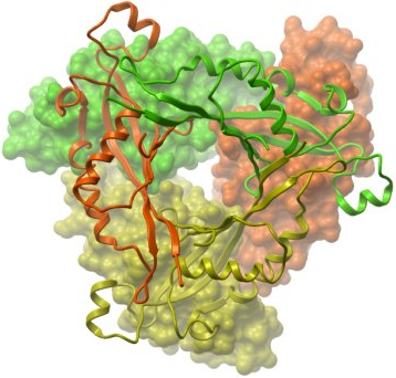
Crystallization: Crystals were grown at 4°C by vapour diffusion in sitting drops by mixing protein (17.75 mg/ml) and well solution containing 20% PEG 3350 and 0.20M Mg formate at a protein to precipitant ratio of 1:1. Crystals were cryo-protected using well solution supplemented with 25% (v/v) ethylene glycol and flash-cooled in liquid nitrogen. |
Data Collection: Resolution: 2.3 Å; X-ray source: SLS-X10. |