RNF128
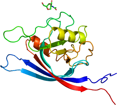
PDB:3ICU
Entry Clone Accession:BC063404
Entry Clone Source:MGC
SGC Clone Accession:grail.038.204.152A03 (SDC152A03)
Tag:N-terminal tag: AAPEHHHHHHDYDIPTTENLYFQGAMD
Sequence: aapehhhhhhdydipttenlyfqgamdGAEAVWTAYLNVSWRVPHTGVNRTVWELSEEGVYGQDSPLEPVAGVLVPPDGPGALNACNPHTNFTVPTVWGSTVQVSWLALIQRGGGCTFADKIHLAYERGASGAVIFNFPGTRNEVIPMSHPGAVDIVAIMIGNLKGTKILQSIQRGIQVTMVIEVGKKHGPWVN
Growth
Procedure: The plasmid grail.038.204.152A03 (SDC152A03) was transformed into DH10Bac E. coli cells (Invitrogen, 10361-012), and a mini-prep was performed to obtain recombinant viral DNA bacmid. SF9 cells (Invitrogen, 11496-015) were transfected with bacmid using Cellfectin reagent (Invitrogen, 10362-010) and recombinant baculovirus was generated. Viral stock was amplified from P1 to P3.
Sf9 cells were grown in Serum Free Medium (HyClone SFX-Insect, SH3027802) at density of 3.5 million cells per milliliter of media and with viability not less then 97 % were infected with 10 mL of P3 viral stock for each 1 L of cell culture. Cell culture medium was collected after 3-4 days of incubation on a shaker at 100 rpm and 27 °C when cells viability dropped to 45-65 %.
Purification
Procedure: A 3.2 L volume of medium was mixed with 30 ml pre-equilibrated NiNTA Superflow beads (Qiagen, 30450) and stirred (Talboys, 134-2) on ice for 1 hour. The resin was transferred to a 100 ml gravity column, washed with 300 ml of Wash Buffer and the protein was eluted with 30-40 ml of Elution Buffer. A second round of NiNTA batch absorption may have been performed for increased yield. The eluate was dialyzed against 50-100 X volume of buffer A overnight at 4 °C. Anion exchange chromatography has been done on HitrapQ HP column (Amersham-Pharmacia, 17-1154-01) using AKTA purifier 100 system. Elution gradient between buffer A and buffer B has been employed. The average yield was about 0.5 mg per liter of insect cell culture. Coomassie-stained SDS-PAGE showed that the product was pure and mass-spectroscopy by LC/MS (Agilent 1100 Series) showed that the protein had the correct molecular weight.
Extraction
Procedure: The cultured medium was centrifuged at 14,000 xg for 15 minutes, and the pH of the supernatant was adjusted to 7.5 at room temperature by adding Titrating Buffer. PMSF (Bioshop, PMS 123.50) and benzamidine (Sigma, B6506-100G) were added to final concentrations of 1 and 2 mM, respectively.
Concentration:Collected protein fractions were concentrated using 15 mL concentrators with an appropriate molecular weight cut-off (Amicon Ultra-15 10,000 MWCO, Millipore, UFC 901024) to a final value of 10 mg/mL.
Structure Determination
Crystallization: Crystals were grown at 18 °C using the hanging drop method in greased VDX Plates (Hampton Research, HR3-306) by mixing equal volumes of protein (9 mg/ml) and Crystallization Buffer (16 % PEG 3350, 0.2 M NaH2PO4 ). Suitable crystals were cryoprotected by immersion in well solution supplemented with 8 % glycerol followed by immersion in 100 % paratone N prior to dunking and storage in liquid nitrogen.
Data Collection:Diffraction data from a crystal of the PA domain was collected on a home-source Rigaku FR-E generator equipped with an R-AXIS IV++ detector and integrated and scaled using the HKL2000 program suite.
Data Processing:The structure was solved by molecular replacement techniques using the program PHASER and truncated and superimposed search models PDB entries 1XF1 and 2EK8. Automated model building using ARP/wARP, combined with iterative model building using the graphics program COOT and maximum-likelihood and TLS refinement with the program REFMAC5 led to a model with an R factor of 19.5% (Rfree 21.8%) for data between 25.0-2.10 Å.