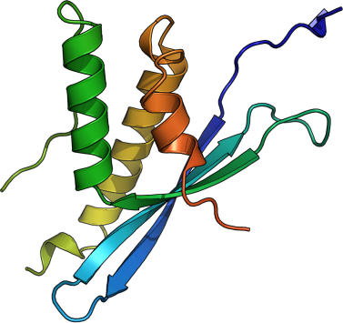
SNX7
PDB:3IQ2
Revision
Revision Type:created
Revised by:created
Revision Date:created
Entry Clone Accession:BC018105
Entry Clone Source:Mammalian Gene Collection (MGC)
SGC Clone Accession:SNX7BA-k046
Tag:C-terminal hexahistidine tag: ahhhhhh
Host:Escherichia coli BL21(DE3) R3 pRARE
Construct
Prelude:Sequence:MDEPDLKDLFITVDEPESHVTTIETFITYRIITKTSRGEFDSSEFEVRRRYQDFLWLKGKLEEAHPTLIIPPLPEKFIVKGMVERFNDDFIETRRKALHKFLNRIADHPTLTFNEDFKIFLTAQAWELSSHahhhhhh
Vector:pNIC-CH
Growth
Medium:Antibiotics:Procedure:Fresh overnight cultures of E. coli strain BL21(DE3) R3 pRARE cells (including 100 µg/ml kanamycin and 34 µg/ml chloramphenicol) transformed with SNX7 expression construct were used to inoculate 0.75 l TB supplemented with 8 g/l glycerol, 50 µg/ml kanamycin and anti-foam PPG P 2000 in a 1 liter flask. Cells were grown in a large scale expression system (Harbinger Biotechnology and Engineering) at 37°C until the OD600 reached ~2.2. The culture was down-tempered to 18°C for 1 h. Expression of SNX7 was induced by adding 0.5 mM IPTG and growth continued over night at 18°C. Cells were harvested by centrifugation at 4400 x g for 10 min. The pellet (20.7g wet cell weight) was resuspended in 50ml lysis buffer supplemented with Complete EDTA-free Protease Inhibitor (Roche Biosciences) and 4 ml benzonase (1000 U). Suspended cells were stored at -80°C until further use.
Purification
ProcedureColumnsIMAC: Ni-charged 1 ml HiTrap Chelating HP (GE Healthcare)
Gel filtration column: HiLoad 16/60 Superdex 75 Prep Grade (GE Healthcare)
Procedure
IMAC columns were equilibrated with IMAC wash1 buffer, and gel filtration columns were equilibrated with GF buffer. Purification of the protein was performed on an ÄKTAxpress system (GE Healthcare). The filtered lysate was loaded onto the Ni-charged HiTrap Chelating column and washed with IMAC wash1 buffer followed by IMAC wash2 buffer. Bound protein was eluted from the IMAC column with IMAC elution buffer and automatically loaded onto the gel filtration column. Fractions containing the target protein were identified by SDS-PAGE, pooled, and fresh TCEP was added to a final concentration of 2 mM. The protein was concentrated using an Amicon Ultra-15 centrifugal filter device (10,000 NMWL; Millipore) to 13.8 mg/ml in a volume of 0.5 ml. The identity of the protein was confirmed by mass spectrometry.
Extraction
ProcedureThe cell suspension was quickly thawed in water. Cells were disrupted by sonication (Vibra-Cell, Sonics) at 80% amplitude for 3 min effective time (pulsed 4s on, 4s off) and cell debris was removed by centrifugation (49000 x g, 20 min, 4 ºC). The supernatant was decanted and filtered through a 0.45 µm flask filter.
Concentration:LigandMassSpec:Crystallization:Crystals were obtained by the sitting drop vapour diffusion method in a 96-well plate. 0.1 µl protein solution (13 mg/ml) including 2 mM Ins(1,3,4,5,6)P5 (C6H17O21P5) was mixed with 0.1 µl of well solution consisting of 0.1 M Sodium Acetate pH 4.6 and 2 M Ammonium Sulfate. The plate was incubated at 4 ºC. Crystals appeared after 28 days and grew to size (approx. 45 µm × 45 µm × 25 µm). The crystals were quickly transferred to a cryo solution consisting of 2.1 M Ammonium Sulfate, 0.1 M Sodium Acetate pH 4.6, 0.3 M NaCl and 20% Glycerol, and flash frozen in liquid nitrogen.
NMR Spectroscopy:Data Collection:Diffraction data to 1.7 Å resolution was collected at MAX-II beamline I911-2.
Data Processing:Data were indexed and integrated in space group P32 using XDS software. The structure was solved by molecular replacement using the structure of the PX-domain of yeast snx3 (pdb: 1ocs) as model template. Chainsaw was used to edit the model and Balbes to solve the structure. The asymmetric unit contained two protein monomers. The space group was P32 with cell dimensions a = b = 57.30Å, c = 97.10Å. Arp/warp was used for initial automatic model building, Refmac5 for refinement and Coot for manual model building. Data in the interval 28.7-1.7 Å resolution were used and refined to R = 16.63% and Rfree = 20.11%. Coordinates for the crystal structure together with structure factors were deposited in the Protein Data Bank, with accession code 3iq2.