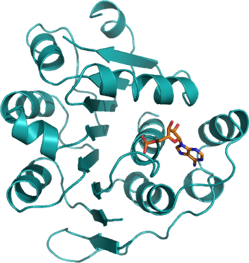
DDX53
PDB:3IUY
Revision
Revision Type:created
Revised by:created
Revision Date:created
Entry Clone Accession:gi|45709415, BC067878
Entry Clone Source:MGC
SGC Clone Accession:DDX53A-s001
Tag:N-terminal hexahistidine tag with integrated TEV protease cleavage site: mhhhhhhssgvdlgtenlyfq*sm
Host:E. coli BL21(DE3) R3 pRARE, where R3 denotes a derivative of BL21(DE3) resistant to a strain of T1 bacteriophage (SGC Oxford) and the pRARE plasmid originating from the Rosetta strain (Novagen) supplies tRNAs for rare codons.
Construct
Prelude:Sequence:MHHHHHHSSGVDLGTENLYFQSMTCDDLKSGEKRLIPKPTCRFKDAFQQYPDLLKSIIRVGILKPTPIQSQAWPIILQGIDLIVVAQTGTGKTLSYLMPGFIHLDSQPISREQRNGPGMLVLTPTRELALHVEAECSKYSYKGLKSICIYGGRNRNGQIEDISKGVDIIIATPGRLNDLQMNNSVNLRSITYLVIDEADKMLDMEFEPQIRKILLDVRPDRQTVMTSATWPDTVRQLALSYLKDPMIVYV
Vector:pNIC-Bsa4
Growth
Medium:Antibiotics:Procedure:Cells from a glycerol stock were grown in 50 mL TB supplemented with 8 g/l glycerol, 100 µg/mL kanamycin and 34 µg/mL chloramphenicol at 37 °C overnight. The overnight culture (50 mL) was used to inoculate 4.5 l TB (divided into 3 x 1.5 l bottles) supplemented with 8 g/l glycerol, 50 µg/mL kanamycin and approximately 200 µl 204 Antifoam A6426 (Sigma) per bottle. The culture was grown in a LEX bioreactor system (Harbinger Biotechnology) at 37 °C until OD600nm had reached 1.6. The culture was down-tempered to 18 °C over a period of 1 hour before target expression was induced by addition of 0.5 mM IPTG. Expression was allowed to continue overnight and cells were harvested the following morning by centrifugation (4,400 x g, 10 min, 4 °C). The resulting cell pellet (110 g wet cell weight) was then stored at -80 °C.
Purification
ProcedureColumnsIMAC: Ni-charged 5 mL HisTrap HP (GE Healthcare)
Gel filtration column: HiLoad 16/60 Superdex 75 Prep Grade (GE Healthcare)
Procedure
The whole purification procedure was performed at 4 ºC. The protein sample was loaded onto an IMAC column previously equilibrated in Lysis buffer. The IMAC column was then washed with, first, 100 mL of IMAC wash1 buffer followed by 100 mL of IMAC wash2 buffer. Bound protein was eluted from the IMAC columns with IMAC elution buffer then loaded onto the gel filtration column equilibrated in GF buffer. Fractions containing the target protein were pooled.
Tag removal
Proteolytic removal of the N-terminal histidine tag was performed. Protein sample was incubated with His-tagged TEV protease in a molar ratio of 30:1 at 4 ºC overnight under agitation. The proteolytic reaction went to completion, as judged by SDS-PAGE. A HisTrap HP column was used to to bind the uncleaved protein fraction. The protein fraction was concentrated in a Vivaspin 20 concentrator (Sartorius) with 10 000 MWCO. The buffer was exchanged to 30 mM Hepes, 500mM NaCl, 10% glycerol, pH 7.5, 2 mM TCEP in this concentrator. Purification Yield: 33 mg DDX53.
Extraction
Procedure1.5 ml Extraction buffer per gram cell pellet and the 1 ml per 1.5 L culture Complete stock solution* was added and the cell pellets were resuspended at 4°C. The resuspended cells were stored in falcon tubes in the -80 freezer. Cells were disrupted by sonication (Vibra-Cell, Sonics) at 80% amplitude for 3 min effective time (pulsed 4s on, 4s off) and cell debris was removed by centrifugation (49,000 x g, 40 min, 4 ºC). The supernatant was decanted and filtered through a 0.45 µm flask filter.
*Complete stock solution: 1 tablet Complete EDTA-free (protease inhibitor) and 8 µL Benzonase (2000 U) dissolved in 1 mL buffer
Concentration:
Ligand
MassSpec:
Crystallization:The Protein solution was incubated with 20 mM ADP and 1 mM MgCl2 for 1 hour. Crystals were obtained by the sitting drop vapour diffusion method in a 96-well plate. 0.1 µl protein solution (16.7 mg/ml mg/mL) was mixed with 0.2 µl of well solution consisting of 0.1M MES, 25% PEG 6000, 0.2 M ammonium chloride. The plate was incubated at 4 ºC and thin plates appeared between 3 and 5 days. The crystals were quickly transferred to a cryo solution consisting of 27% PEG6000, 0.1M MES pH 6, 0.2M ammonium chloride, 15% glycerol, 0.2M NaCl and flash frozen in liquid nitrogen.
NMR Spectroscopy:
Data Collection:Diffraction data to 2.4 Å resolution was collected at ESRF beamline ID14-2.
Data Processing:Intensity integration was performed by MOSFLM. Two datasets were combined and then scaled with SCALA. The space group was P21 with cell dimensions a = 56.4 Å, b = 61.3 Å c = 65.8 Å, α = 90°, β = 96.4°, γ = 90°. The structure was solved by molecular replacement using MOLREP with the structure of human DEAD-BOX RNA helicase DDX5 as template (PDB: 3FE2). Structure refinement was performed with REFMAC 5.5.0035. R-factor = 0.20, R-free = 0.25.