ACOT4A
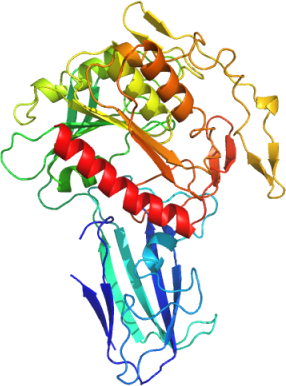
PDB:3K2I
Revision
Revision Type:created
Revised by:created
Revision Date:created
Entry Clone Accession:BC090945
Entry Clone Source:Mammalian Gene Collection
SGC Clone Accession:ACOT4A-k012
Tag:N-terminal hexahistidine tag with integrated TEV protease cleavage site: mhhhhhhssgvdlgtenlyfq*sm
Host:E.coli Rosetta 2 (Novagen)
Construct
Prelude:Sequence:mhhhhhhssgvdlgtenlyfq*smSATLILEPPGRCCWNEPVRIAVRGLAPEQRVTLRASLRDEKGALFRAHARYCADACGELDLERAPALGGSFAGLEPMGLLWALEPEKPFWRFLKRDVQIPFVVELEVLDGHDPEPGRLLCQAQHERHFLPPGV
WRQSVRAGRVRATLFLPPGPGPFPGIIDIFGIGGGLLEYRASLLAGHGFATLALAYYNFEDLPNNMDNISLEYFEEAVCYMLQHPQVKGPGIGLLGISLGADICLSMASFLKNVSATVSINGSGISGNTAINYKHSSIPPLGYDLRRIKVAFSGLVDIVDIRNALVGGYKNPSMIPIEKAQGPILLIVGQDDHNWRSELYAQTVSERLQAHGKEKPQIICYPGTGHYIEPPYFPLCPASLHRLLNKHVIWGGEPRAHSKAQEDAWKQILAFFCKHLGGTQKTAVPKL
W in red bold indicates position of R-to-W mutation revealed by sequencing
Vector:pNIC-Bsa4
Growth
Medium:Antibiotics:Procedure:Cells from a glycerol stock were used to inoculate 40 ml LB supplemented with 50 µg/ml kanamycin and 34 μg/ml chloramphenicol, and grown at 30 °C overnight. 2 x 15 ml of the overnight culture was used to inoculate 2 x 1.5 l TB supplemented with 8 g/l glycerol, 50 µg/ml kanamycin and approximately 5-10 drops of Dow Corning Antifoam. The culture was grown in a LEX bioreactor system (Harbinger Biotechnology) at 37 °C until OD600 reached ~2. The culture was down-tempered to 18 °C over a period of one hour before target expression was induced by addition of 0.5 mM IPTG. Expression was allowed to continue overnight and cells were harvested the following morning by centrifugation (4,400 x
g, 10 min, 4 °C). The resulting cell pellet (69.2 g wet cell weight) was resuspended in lysis buffer (2 ml/g cell pellet) supplemented with one tablets of Complete EDTA-free protease inhibitor (Roche Applied Science) and 2000 U Benzonase (Merck) per 100 ml lysis buffer, and stored at -80 °C.
Purification
ProcedureColumnsIMAC: Ni-charged 5 ml HiTrap Chelating HP (GE Healthcare)
Gel filtration column: HiLoad 16/60 Superdex 200 Prep Grade (GE Healthcare)
Procedure
Purification of the native protein was performed as a two step process on an ÄKTAxpress system (GE Healthcare). Prior to purification, columns were equilibrated with IMAC wash1 buffer and gel filtration buffer, respectively. The filtered lysate was loaded onto the Ni-charged HiTrap Chelating column and washed with IMAC wash1 buffer followed by IMAC wash2 buffer. Bound protein was eluted from the IMAC column with IMAC elution buffer and automatically loaded onto the gel filtration column. Fractions containing the target protein were pooled and subsequently concentrated using an Amicon Ultra-15 centrifugal filter device, 30,000 NMWL (Millipore). The concentration was measured to 14.5 mg/ml in a volume of 0.2 ml and the samples were stored at -80 ºC. The identity of the protein was confirmed by mass spectrometry.
Extraction
ProcedureThe cell suspension was quickly thawed in water. Cells were disrupted by sonication (VibraCell, Sonics) at 80% amplitude for 3 min effective time (puls 4s on, 4s off) and cell debris was removed by centrifugation (49,000 x
g, 30 min, 4 ºC). The supernatant was decanted and filtered through a 0.45 µm flask filter.
Concentration:LigandMassSpec:Crystallization:Native crystals were obtained by the sitting drop vapour diffusion method in a 96-well plate. 0.2 µl of the protein solution (14.0 mg/ml) including 2.5 mM acetyl CoA was mixed with 0.1 µl of well solution consisting of 0.2M sodium iodide, 0.1M Bis-Tris propane pH 8.5, 20% (w/v) PEG 3350. The plate was incubated at 4 ºC and crystals were obtained after 5 days. The crystals were quickly transferred to cryo solution containing well solution and 20% glycerol, and flash frozen in liquid nitrogen.
NMR Spectroscopy:Data Collection:Native data was collected to 2.4 Å at DIAMOND (I02). The crystal belonged to space group P 21 21 21 with cell parameters of a= 72.47 Å b=93.00 Å and c=133.72Å.
Data Processing:Data integration was performed in imosflm and scaled with scala. Molecular replacement was done using molrep with model 3HLK. The asymmetric unit consisted of two chains. The structure was refined with Refmac5 with a final refinement cycle using phenix.refine. Final R-values were R= 19.25% and Rfree= 24.89% and coordinates and structure factors were deposited in the PDB with accession code 3K2I.