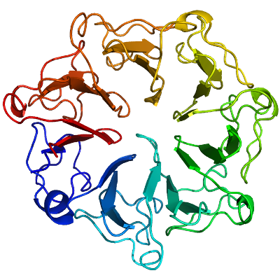
HERC2
PDB:3KCI
Entry Clone Accession:Herc2.LIFESEQ3382404.OBS.IHS1382-8549203.pINCY
Entry Clone Source:OpenBiosystems
SGC Clone Accession:herc2.3951.4321 (SDC174D02)
Tag:N-terminal tag: MHHHHHHSSGRENLYFQG
Vector:pET28a-LIC
Sequence: mhhhhhhssgrenlyfqgSGTIYGWGHNHRGQLGGIEGAKVKVPTPCEALATLRPVQLIGGEQTLFAVTADGKLYATGYGAGGRLGIGGTESVSTPTLLESIQHVFIKKVAVNSGGKHCLALSSEGEVYSWGEAEDGKLGHGNRSPCDRPRVIESLRGIEVVDVAAGGAHSACVTAAGDLYTWGKGRYGRLGHSDSEDQLKPKLVEALQGHRVVDIACGSGDAQTLCLTDDDTVWSWGDGDYGKLGRGGSDGCKVPMKIDSLTGLGVVKVECGSQFSVALTKSGAVYTWGKGDYHRLGHGSDDHVRRPRQVQGLQGKKVIAIATGSLHCVCCTEDGEVYTWGDNDEGQLGDGTTNAIQRPRLVAALQGKKVNRVACGSAHTLAWSTSKP
Growth
Procedure: Competent BL-21(DE3) cells (Invitrogen, C6000-03) were transformed and grown using the LEX system (Harbinger BEC) at 37 degC in 2L bottles (VWR, 89000-242) containing 1800 ml of TB (Sigma T0918) supplemented with 150 mM glycerol, 100 µM Kanamycin and 600 µl antifoam 204 (Sigma A-8311). At OD600 = 6, the temperature was reduced to 15 degC, and one hour later the culture was induced with 100 µM IPTG (BioShop IPT001) and incubated overnight (16 hours) at 15 degC. Cell pellets were collected by centrifugation (12,227 xg, 20 mins) and frozen in liquid nitrogen.
Purification
Procedure:
Cleared lysate was rocked with TALON metal-affinity resin (BD Biosciences) (1.5 mL settled beads per L cell culture) at 4 °C. Resin was transferred to a column and washed with 5 column volumes (cv) of Wash buffer A, 5 cv of Wash buffer B, and 5 cv of Wash buffer A. The protein was eluted with 2 cv of Elution buffer. The protein was further purified by gel filtration through a HighLoad 16/60 Superdex 200 column (GE Healthcare) equilibrated with Gel Filtration buffer. Fractions containing protein (analyzed by ABS280 nm) were pooled and concentrated to 0.5-1.0 mM using concentrators (Amicon) with 5 kDa cutoff. The yield of the protein was approximately 10 mg per L of bacterial culture. Coomassie-stained, SDS-PAGE showed that the product was pure and Mass-spectroscopy by LCMS (Agilent 1100 Series) showed that the protein has 107 Da more than the calculated molecular weight.
Extraction
Procedure:
Cell pellets were resuspended in Lysis buffer(30 mL per L culture), lysed using a Microfluidizer (Microfluidics, M110-EH) at 18,000 psi, and cleared by centrifugation (40,000 xg for 30 minutes).
Structure Determination
Crystallization:Crystals were grown at 18 oC using the sitting drop method in 96well plates (Art Robbins, 102-0004-00) by mixing equal volumes of protein (8.0 mg/ml) and Crystallization Buffer (20% PEG3350, 0.2mM MgCl2). Suitable crystals were cryoprotected by immersion in well solution supplemented with 25% (v/v) Glycerol prior to dunking and storage in liquid nitrogen.
Data Collection:Diffraction data from a crystal of the third RCC1-like domain of HERC2 was collected on a home source Rigaku FR-E, and integrated and scaled using the HKL2000 program suite.
Data Processing:The structure was solved by molecular replacement techniques using the program PHASER and search model PDB entry 1A12. Automated model building using ARP/wARP, combined with iterative model building using the graphics program Coot and maximum-likelihood and TLS refinement with the program REFMAC5 led to a model with an R factor of 15.2% (Rfree 19.5%) for data between 20.0-1.8 Å. Parameters for Translation/liberation/screw (TLS) refinement were generated using the TLSMD web server.