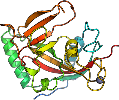
Tankyrase-2
PDB:3KR7
Revision
Revision Type:created
Revised by:created
Revision Date:created
Entry Clone Accession:NM_025235
Entry Clone Source:Origene
SGC Clone Accession:TNKS2A-s001
Tag:N-terminal hexahistidine tag: MHHHHHHSSGVDLGTENLYFQSM
Host:Escherichia coli BL21(DE3) R3 pRARE
Construct
Prelude:Sequence:MHHHHHHSSGVDLGTENLYFQSMLNTSGSGTILIDLSPDDKEFQSVEEEMQSTVREHRDGGHAGGIFNRYNILKIQKVCNKKLWERYTHRRKEVSEENHNHANERMLFHGSPFVNAIIHKGFDERHAYIGGMFGAGIYFAENSSKSNQYVYGIGGGTGCPVHKDRSCYICHRQLLFCRVTLGKSFLQFSAMKMAHSPPGHHSVTGRPSVNGLALAEYVIYRGEQAYPEYLITYQIMRPEG
Vector:pNIC-Bsa4
Growth
Medium:Fresh overnight cultures of E. coli strain BL21(DE3) R3 pRARE cells (including 100 µg/ml kanamycin and 34 µg/ml chloramphenicol) transformed with TNKS2 expression construct were used to inoculate 4.5 l TB supplemented with 8 g/l glycerol, 50 µg/ml kanamycin and anti-foam PPG P2000 in three 2-liter flasks. Cells were grown in a large scale expression system (Harbinger Biotechnology and Engineering) at 37°C until the OD600 reached ~1.6. The culture was down-tempered to 18°C for 1 h. Expression of TNKS2 was induced by adding 0.5 mM IPTG and growth continued over night at 18°C. Cells were harvested by centrifugation at 4400 x g for 10 min. The pellet (29g wet cell weight) was resuspended in 90ml lysis buffer supplemented with Complete EDTA-free Protease Inhibitor (Roche Biosciences) and benzonase Suspended cells were stored at -80°C until further use.
Antibiotics:Procedure:Purification
ProcedureColumnsIMAC: Ni-charged 1 ml HiTrap Chelating HP (GE Healthcare)
Gel filtration column: HiLoad 16/60 Superdex 75 Prep Grade (GE Healthcare)
Procedure
Purification of the protein was performed on an ÄKTAxpress system (GE Healthcare). Prior to purification, IMAC column was equilibrated with IMAC wash1 buffer and gel filtration column with gel filtration buffer. The filtered lysate was loaded onto the IMAC column, and therafter washed with IMAC wash1 buffer followed by IMAC wash2 buffer. Bound protein was eluted from the IMAC column with IMAC elution buffer and automatically loaded onto the gel filtration column. Eluting fractions were analyzed by SDS-PAGE and target protein was pooled. Fresh TCEP was added to a final concentration of 2 mM. Purified protein was difficult to concentrate using an Amicon Ultra-15 centrifugal filter device (Millipore, 10,000 MWCO). Chymotrypsin (1:100 w/w) was added and the protein was concentrated to 13.7 mg/ml. Mass spectrometry, identified two chymotrypsin cleaved TNKS2 fragments.
Extraction
ProcedureThe cell suspension was quickly thawed in water. Cells were disrupted by sonication (Vibra-Cell, Sonics) at 80% amplitude for 3 min effective time (pulsed 4s on, 4s off) and cell debris was removed by centrifugation (49000 x g, 20 min, 4 ºC). The supernatant was decanted and filtered through a 0.45 µm flask filter.
Concentration:LigandMassSpec:Crystallization:Crystals were obtained by the sitting drop vapour diffusion method in a 96-well plate. 0.1 µl protein solution (13.7 mg/ml) was mixed with 0.2 µl of well solution consisting of 15% PEG3350, 0.2M lithium sulfate, 0.1M Tris-HCl pH 8.5. The plate was incubated at 4 ºC. Crystals grew in 4 days wherafter they were quickly transferred to a cryo solution consisting of 17% PEG3350, 0.2M lithium sulfate, 0.1M Tris-HCl pH 8.5, 25% Glycerol, 0.2M NaCl and flash frozen in liquid nitrogen.
NMR Spectroscopy:Data Collection:Diffraction data to 1.95 Å resolution was collected at DIAMOND beamline I03.
Data Processing:Data were indexed and integrated in space group P41212 using XDS software. The structure was solved by molecular replacement using the structure of the catalytic domain of the previously determined human tankyrase-1 (pdb: 2rf5) as model template. Molrep was used to solve the structure. The asymmetric unit contained one protein monomer. The cell dimensions are a = 66.9Å, b = 66.9Å, c = 118.1Å. Refmac5 was used for refinement and Coot for model building. Data in the interval 45.0-1.95 Å resolution were used and refined to R = 18.09% and Rfree = 21.85%. Coordinates for the crystal structure were deposited in the Protein Data Bank, with accession code 3kr7.