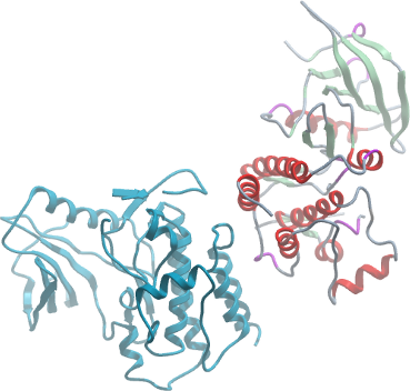
EPHA8
PDB:3KUL
Revision
Revision Type:created
Revised by:created
Revision Date:created
Entry Clone Accession:epha8.NM_020526.HIP.HsCD00080451.pENTR223.1
Entry Clone Source:Harvard Institute of Proteomics
SGC Clone Accession:epha8.0603.0909173A11 (SDC173A11)
Tag:N-terminal tag: MHHHHHHSSGRENLYFQG
Host:BL21 (DE3)
Construct
Prelude:
Sequence:mhhhhhhssgrenlyfqgKLPEPQFYAEPHTYEEPGRAGRSFTREIEASRIHIEKIIGSGDSGEVCYGRLRVPGQRDVPVAIKALKAGYTERQRRDFLSEASIMGQFDHPNIIRLEGVVTRGRLAMIVTEYMENGSLDTFLRTHDGQFTIMQLVGMLRGVGAGMRYLSDLGYVHRDLAARNVLVDSNLVCKVSDFGLSRVLEDDPDAAYTTTGGKIPIRWTAPEAIAFRTFSSASDVWSFGVVMWEVLAYGERPYWNMTNRDVISSVEEGYRLPAPMGCPHALHQLMLDCWHKDRAQRPRFSQIVSVLDALIRSPESLRATATVS
Vector:pET28-MHL
Growth
Medium:TB
Antibiotics:
Procedure:Competent BL21 (DE3) cells (Invitrogen, C6000-03) were transformed and grown using the LEX system (Harbinger BEC) at 37 °C in 1L bottles (VWR, 89000-242) containing 900 ml of TB (Sigma, T0918) supplemented with 150 mM glycerol, 100 µM Kanamycin, and 600 µl antifoam 204 (Sigma, A-8311). At OD600 = 6, temperature was reduced to 15 °C, and one hour later the culture was induced with 100 µM IPTG (BioShop, IPT001). Cultures were grown overnight (16 hours) at 15 °C, and cell pellets were collected by centrifugation (12,227 xg, 20 mins) and frozen at -80 degC.
Purification
Procedure
The cleared lysate was mixed with HisLink (Promega, V882A), 2.0 mL settled resin per 40 mL lysate for 60 minutes at 4 °C. The resin was spun (500 xg for 2 minutes), batch-washed with 2X45 mL of cold Wash Buffer, and transferred to a column. After additional washing (50 column volumes), protein was eluted with 40 mL of Elution Buffer and dialyzed against 50 volumes of Dialyses Buffer overnight at 4 °C. The protein sample was concentrated using concentrators with an appropriate molecular weight cut-off (Amicon Ultra-15 10,000 MWCO, Millipore, UFC 901024) to a final value of 20 mg/mL. Protein yield was 13 mg per liter of bacterial culture. Coomassie-stained SDS-PAGE showed that the product was pure.
Extraction
Procedure
After resuspension with an Ultra-Turrax T18 homogenizer (IKA Works) in 40 mL per liter bacterial culture of Lysis Buffer, cells were lysed by sonication (Misonix 3000, 15-338-276) on ice for 10 minutes total sonication time (10 sec pulses at half-maximal frequency with 10 second rest).
Concentration:
Ligand
MassSpec:Mss-spectroscopy by LCMS (Agilent 1100 Series) showed that the purified protein was slightly larger than expected and somewhat heterogeneous.
Crystallization:Crystals were grown at 18 °C using the sitting drop method in 96-2-well plates (Hampton Research, HR 3-299) by mixing equal volumes of protein (20 mg/ml; 0.54 mM) and Crystallization Buffer (20 % PEG 8000, 0.2 M (NH4)2SO4, 0.1 M HEPES pH7.5). Immediately prior to setting-up crystallization plates, chymotrypsin was added to protein samples to a final concentration of 5.7e-7 M (0.57 µM). Crystals were harvested without cryoprotection into liquid nitrogen.
NMR Spectroscopy:
Data Collection:Diffraction data from a crystal of the kinase domain was collected at beamline 23 ID-B at the Argonne Photon Source. The data set was integrated and scaled using the HKL2000 program suite.
Data Processing:The structure was solved by molecular replacement techniques using the program PHASER and search model PDB entry 2REI. Automated model building using ARP/wARP, combined with iterative model building using the graphics program Coot and maximum-likelihood and TLS refinement with the program REFMAC5 led to a model with an R factor of 18.7% (Rfree 24.4%) for data between 50-2.15 Å. Parameters for Translation/liberation/screw (TLS) refinement were generated using the TLSMD web server.