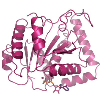
DHX9
PDB:3LLM
Revision
Revision Type:created
Revised by:created
Revision Date:created
Entry Clone Accession:gi|4503297
Entry Clone Source:TC109149/NM001357
SGC Clone Accession:DHX9A-s001
Tag:N-terminal hexahistidine tag with integrated TEV protease cleavage site: mhhhhhhssgvdlgtenlyfq*sm
Host:E.coli BL21(DE3) R3 pRARE supplies tRNAs for rare codons.
Construct
Prelude:Sequence:MHHHHHHSSGVDLGTENLYFQSMNQVGVVPWSPPQSNWNPWTSSNIDEGPLAFATPEQISMDLKNELMYQLEQDHDLQAILQERELLPVKKFESEILEAISQNSVVIIRGATGCGKTTQVPQFILDDFIQNDRAAECNIVVTQPRRISAVSVAERVAFERGEEPGKSCGYSVRFESILPRPHASIMFCTVGVLLRKLEAGIRGISHVIVDEIHERDINTDFLLVVLRDVVQAYPEVRIVLMSATIDTSMFCEYFFNCPIIEV
Vector:pNIC-Bsa4
Growth
Medium:Antibiotics:Procedure:Cells from a glycerol stock were grown in 50 mL TB supplemented with 8 g/l glycerol, 100 µg/mL kanamycin and 34 µg/mL chloramphenicol at 37 °C overnight. The overnight culture (50 mL) was used to inoculate 1.5 l TB (divided into 2 x 0.75 l bottles) supplemented with 8 g/l glycerol, 50 µg/mL kanamycin and approximately 200 µl 204 Antifoam A6426 (Sigma) per bottle. The culture was grown in a LEX bioreactor system (Harbinger Biotechnology) at 37 °C until OD600nm had reached 1-2. The culture was down-tempered to 18 °C over a period of 1 hour before target expression was induced by addition of 0.5 mM IPTG. Expression was allowed to continue overnight and cells were harvested the following morning by centrifugation (4,400 x g, 10 min, 4 °C). The resulting cell pellet (30 g wet cell weight) was then stored at -80 °C.
Purification
ProcedureColumnsIMAC: Ni-charged 5 mL HisTrap HP (GE Healthcare)Gel filtration column: HiLoad 16/60 Superdex 75 Prep Grade (GE Healthcare)
Procedure
The whole purification procedure was performed at 4 ºC. The protein sample was loaded onto an IMAC column previously equilibrated in Lysis buffer. The IMAC column was then washed with, first, 100 mL of IMAC wash1 buffer followed by 100 mL of IMAC wash2 buffer. Bound protein was eluted from the IMAC columns with IMAC elution buffer then loaded onto the gel filtration column equilibrated in GF buffer. Fractions containing the target protein were pooled.
Tag removal:
Proteolytic removal of the N-terminal histidine tag was performed. Protein sample was incubated with His-tagged TEV protease in a molar ratio of 50:1at 4 ºC at 4 ºC overnight under agitation. The proteolytic reaction went to completion, as judged by SDS-PAGE. A HisTrap HP column was used to to bind the unvleaved protein fraction. The protein was concentrated in a Vivaspin 20 concentrator (Sartorius) with 10 000 MWCO. The buffer was exchanged in the concentrator to: 20 mM HEPES, 300 mM NaCl, 10% glycerol, 0.5 mM TCEP. Yield: 36 mg Protein.
Extraction
ProcedureCells were harvested by centrifugation and the cell pellets were resuspended in 1.5 volumes/wet cell weight of lysis buffer (100 mM HEPES, 500 mM NaCl, 10% glycerol, 10 mM imidazole, 0.5 mM TCEP, pH 8.0, and one tablet of Complete EDTA-free protease inhibitor (Roche Biosciences) per 50 ml cell suspension). Before lysis, 4 µl (1000 U) of Benzonase (Novagen) was added per 50 ml cell suspension, and lysis was achieved by sonication (Vibra-Cell, Sonics, at 80% amplitude for 3 min effective time, pulsed 4s on, 4s off). Cell debris was removed by centrifugation (49,000 x g, 40 min, 4 ºC) and the soluble fractions were filtered through a syringe filter (0.45 μm pore size). Selenomethionine-labelled protein was produced by the pathway-inhibition method (Van Duyne, Standaert et al. 1993).
Concentration:LigandMassSpec:Crystallization:The Protein solution was incubated with 1 mM ADP and 1 mM MgCl2 for 1 hour. Crystals were obtained by the sitting drop vapour diffusion method in a 96-well plate. 0.3 µl protein solution (20 mg/ml) was mixed with 0.2 µl of well solution consisting of 160 mM calcium acetate hydrate, 80 mM sodium cacodylate pH 6.5, 14.4% w/v PEG 8000, 20% glycerol. The plate was incubated at 4 ºC and crystals appeared after 3-5 days. The crystals were transferred to a cryo solution consisting of 128 mM calcium acetate hydrate, 64 mM sodium cacodylate pH 6.5, 13% w/v PEG 8000, 30% glycerol and flash frozen in liquid nitrogen.
NMR Spectroscopy:Data Collection:Diffraction data to 2.8 Å resolution was collected at Diamond beamline I03.
Data Processing:A 3-wavelength anomalous dispersion (MAD) experiment was performed. The data were integrated with Mosflm and scaled with Scala. The space group was P3221 with cell dimensions a = 113.7, b = 113.7, c = 141.65, alpha = 90°, beta = 90°, gamma = 120°. Selen positions were determined by SHELXD (Sheldrick 2008). Phases were computed and improved with autoSHARP (Vonrhein, Blanc et al. 2007). Model building was performed by Buccaneer (Cowtan 2006) and Coot (Emsley and Cowtan 2004). Refinement was effected using REFMAC (Murshudov, Vagin et al. 1997). R-factor = 0.211, R-free = 0.238.