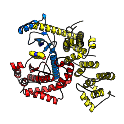
PIK3C3
PDB:3LS8
Revision
Revision Type:created
Revised by:created
Revision Date:created
Entry Clone Accession:BC033004
Entry Clone Source:Mammalian Gene Collection
SGC Clone Accession:PIK3C3A-k016
Tag:N-terminal hexahistidine tag with integrated TEV protease cleavage site: mhhhhhhssgvdlgtenlyfq*sm
Host:E.coli BL21(DE3) R3 pRARE, where R3 denotes a derivative of BL21(DE3) resistant to a strain of T1 bacteriophage (SGC Oxford) and the pRARE plasmid originating from the Rosetta strain (Novagen) supplies tRNAs for rare codons.
Construct
Prelude:Sequence:mhhhhhhssgvdlgtenlyfq*smSKHHKLARSLRSGPSDHDLKPNAATRDQLNIIVSYPPTKQLTYEEQDLVWKFRYYLTNQEKALTKFLKCVNWDLPQEAKQALELLGKWKPMDVEDSLELLSSHYTNPTVRRYAVARLRQADDEDLLMYLLQLVQALKYENFDDIKNGLEPTKKDSQSSVSENVSNSGINSAEIDSSQIITSPLPSVSSPPPASKTKEVPDGENLEQDLCTFLISRACKNSTLANYLYWYVIVECEDQDTQQRDPKTHEMYLNVMRRFSQALLKGDKSVRVMRSLLAAQQTFVDRLVHLMKAVQRESGNRKKKNERLQALLGDNEKMNLSDVELIPLPLEPQVKIRGIIPETATLFKSALMPAQLFFKTEDGGKYPVIFKHGDDLRQDQLILQIISLMDKLLRKENLDLKLTPYKVLATSTKHGFMQFIQSVPVAEVLDTEGSIQNFFRKYAPSENGPNGISAEVMDTYVKSCAGYCVITYILGVGDRHLDNLLLTKTGKLFHIDFGYILGRDPKPLPPPMKLNKEMVEGMGGTQSEQYQEFRKQCYTAFLHLRRYSNLILNLFSLMVDANIPDIALEPDKTVKKVQDKFRLDLSDEEAVHYMQSLIDESVHALFAAVVEQIH
Vector:pNIC-Bsa4
Growth
Medium:Antibiotics:Procedure:Cells from a glycerol stock were grown in 20 ml TB supplemented with 8 g/l glycerol, 100 µg/ml kanamycin and 34 µg/ml chloramphenicol at 30 °C overnight. 15 ml of the overnight culture was used to inoculate 750 ml TB supplemented with 8 g/l glycerol, 50 µg/ml kanamycin and approximately 200 µl 204 Antifoam A6426 (Sigma). The culture was grown in a LEX bioreactor system (Harbinger Biotechnology) at 37 °C until OD600 reached ~2. The culture was down-tempered to 18 °C over a period of 1 hour before target expression was induced by addition of 0.5 mM IPTG. Expression was allowed to continue overnight and cells were harvested the following morning by centrifugation (4,430 x
g, 10 min, 4 °C). The resulting cell pellet (13 g wet cell weight) was resuspended in lysis buffer (1.5 ml/g cell pellet), supplemented with 1000 U Benzonase (Merck) and half a tablet of Complete EDTA-free protease inhibitor (Roche Applied Science). The cell suspension was stored at -80 °C.
Purification
ProcedureColumnsIMAC: Ni-charged 1 ml HiTrap Chelating HP (GE Healthcare)
Gel filtration column: HiLoad 16/60 Superdex 200 Prep Grade (GE Healthcare)
Procedure
IMAC columns were equilibrated with IMAC wash1 buffer, and gel filtration columns were equilibrated with GF buffer. Purification of the protein was performed on an ÄKTAxpress system (GE Healthcare). The filtered lysate was loaded onto the Ni-charged HiTrap Chelating column and washed with IMAC wash1 buffer followed by IMAC wash2 buffer. Bound protein was eluted from the IMAC column with IMAC elution buffer and automatically loaded onto the gel filtration column. Fractions containing the target protein were identified by SDS-PAGE, pooled, and fresh TCEP was added to a final concentration of 2 mM. The protein was concentrated using an Amicon Ultra-15 centrifugal filter device (30,000 NMWL; Millipore) to 30 mg/ml in a volume of 0.5 ml.
Tag removal
The N-terminal histidine tag was proteolytically removed by incubating the target protein with His-tagged TEV protease in a molar ratio of 50:1 at 4 ºC overnight. Then, additional TEV-protease was added to a molar ratio of 25:1 and the reaction was performed at RT during two more hours. The proteolytic reaction went to completion, as judged by SDS-PAGE. The sample was loaded onto a Ni-charged 1 ml HiTrap Chelating HP column (GE Healthcare) pre-equilibrated with IMAC wash1 buffer. The column was then washed in IMAC wash2 buffer. Fractions corresponding to the flowthrough and the washing step were concentrated to 0.5 ml in a Vivaspin 20 ml (30,000 NMWL; Sartorius), diluted to 3 ml in TEV purification buffer, then loaded onto two Ni-charged 1ml HiTrap Chelating HP columns connected in series equilibrated in TEV purification buffer. The columns were then washed in IMAC wash1 buffer followed by a wash in IMAC wash2 buffer. The cleaved protein eluted in the IMAC wash2 buffer. The fractions containing the cleaved protein were pooled and concentrated in a Vivaspin 20 ml (30,000 NMWL). The sample was diluted in GF buffer then concentrated in a Vivaspin 20 ml (30,000 NMWL). The final protein concentration was determined to 20.8 mg/ml in a volume of 0.35 ml The identity of the protein was confirmed by mass spectrometry.
Extraction
ProcedureThe cell suspension was quickly thawed in water. Cells were disrupted by sonication (Vibra-Cell, Sonics) at 80% amplitude for 3 min effective time (pulsed 4s on, 4s off) and cell debris was removed by centrifugation (49,000 x
g, 20 min, 4 ºC). The supernatant was decanted and filtered through a 0.45 µm flask filter.
Concentration:LigandCompound 15E
MassSpec:Crystallization:Compound 15 E was added to the protein solution to a final concentration of 1mM. Sample is then spinned at 24100 x g for 5 min and supernatant is used to set-up crystallization plates. Crystals were obtained by the sitting drop vapour diffusion method in a 96-well plate. 0.1 µl of the mix between protein solution (20.6 mg/ml) and Compound 15 E (1mM) was mixed with 0.2 µl of well solution consisting of 0.1 M Hepes pH 7.5, 25% PEG 3350 and 0.2 M ammonium acetate. The plate was incubated at 20 ºC and crystals appeared within 5 days. The crystals were quickly transferred to a cryo solution consisting of 50 mM Hepes pH 7.5, 26% PEG3350, 0.2 M Ammonium acetate, 0.3M NaCl, 2mM Compound 15 E, 20% glycerol and flash frozen in liquid nitrogen.
NMR Spectroscopy:Data Collection:Diffraction data to 2.25 Å resolution was collected at DIAMOND, beamline I04.
Data Processing:The structure was solved by molecular replacement using apo PIK3C3 as template (PDB: 3IHY). The space group was P212121 with cell dimensions a=61.686 Å b=141.141 Å c=163.975 Å. Two monomers were located by MOLREP in the asymmetric unit. Compound 15 E was located manually in the Fo-Fc difference map. REFMAC was used for refinement and Coot for model building. TLS refinement using 4 TLS groups per chains was used at the end of the refinement process. Data in the interval 70.9-2.25 Å resolution was used and at the end of the refinement the R values were: R=21.1% and Rfree=25.1%. Coordinates for the crystal structure were deposited in the Protein Data Bank, accession code 3LS8.