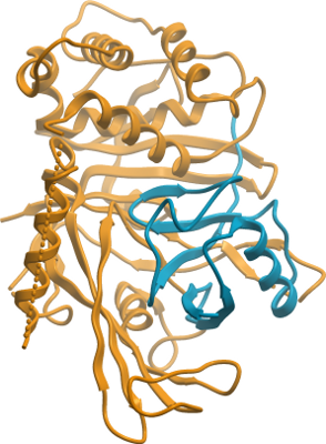
USP21
PDB:3MTN
Entry Clone Accession:usp21.LIFESEQ2303317.OBS.IHS1382-8528801.pSPORT
Entry Clone Source:Open Biosystems
SGC Clone Accession:usp21.209-562.122G07 (SDC122G07)
Tag:N-terminal tag: MGSSHHHHHHSSGLVPRGS
Host:Competent BL21 (DE3) cells (Invitrogen C6000-03)
Vector:pET28a-LIC vector (GenBank EF442785)
Sequence: SGHVGLRNLGNTCFLNAVLQCLSSTRPLRDFCLRRDFRQEVPGGGRAQELTEAFADVIGALWHPDSCEAVNPTRFRAVFQKYVPSFSGYSQQDAQEFLKLLMERLHLEINRRGRRAPPILANGPVPSPPRRGGALLEEPELSDDDRANLMWKRYLEREDSKIVDLFVGQLKSCLKCQACGYRSTTFEVFCDLSLPIPKKGFAGGKVSLRDCFNLFTKEEELESENAPVCDRCRQKTRSTKKLTVQRFPRILVLHLNRFSASRGSIKKSSVGVDFPLQRLSLGDFASDKAGSPVYQLYALCNHSGSVHYGHYTALCRCQTGWHVYNDSRVSPVSENQVASSEGYVLFYQLMQEPP
Growth
Medium:TB (Sigma T0918) supplemented with 150 mM glycerol, 100 µM Kanamycin and 600 µl antifoam 204 (Sigma A-8311)
Procedure:Competent BL21 (DE3) cells (Invitrogen C6000-03) were transformed and grown using the LEX system (Harbinger BEC) at 37 °C in 2L bottles (VWR 89000-242) containing 1800 ml of TB (Sigma T0918) supplemented with 150 mM glycerol, 100 µM Kanamycin and 600 µl antifoam 204 (Sigma A-8311). When OD600 ~ 6 was reached, the temperature was reduced to 15 °C, and one hour later protein expression was induced with 100 µM IPTG (BioShop IPT001) and the culture was incubated overnight (16 hours) at 15 °C. Cell pellets were collected by centrifugation (12,227 xg, 20 min), frozen and stored at -80 °C.
Purification
Procedure: The cleared lysate was loaded onto a 3 mL TALON metal-affinity resin column (BD Biosciences) at 4ºC. The column was washed with 10 mL Wash Buffer A, 10 mL Wash Buffer B and 10 mL Wash Buffer A. The protein was eluted with 6 mL Elution Buffer. The protein was further purified by gel filtration on a HighLoad 16/60 Superdex 200 column (GE Healthcare) using Gel Filtration Buffer. Fractions containing protein corresponding to the USP21 peak were pooled and concentrated by ultrafiltration. The yield of the protein was 6 mg per litre bacterial culture.
Extraction
Procedure: After resuspension in 30 mL per litre bacterial culture of Lysis Buffer, cells were lysed using a Microfluidics M110-EH microfluidizer at 18,000 psi.
Structure Determination
Crystallization: USP21 solution (500 µl, 2.2 mg/ml) was mixed with Ubv.21.4 (100 µl, 4.9 mg/ml), which resulted in USP21:Ubv.21.4 molar ratio of 1:1.6. The mixture was incubated for 1 h at ambient temperature (294 K) followed by incubation for 16 h at 281 K and concentrated by ultrafiltration to a final volume of 150 µl, which resulted in USP21 concentration of 9 mg/ml. Crystals of the USP21-inhibitor complex were grown at 291 K using the hanging drop method by mixing equal volumes of the above complex solution and Crystallization Buffer (11% polyethyleneglycol 4000, 0.1 M sodium citrate, pH 5.3, 0.1 M ammonium acetate and 0.5 mM TCEP). The crystals were cryoprotected by immersion in the Crystallization Buffer supplemented with 25% (v/v) glycerol and placed in liquid nitrogen.
Data Processing:Diffraction data from a crystal of the USP21 catalytic domain in complex with Ubv.21.4 inhibitor was collected on a home-source Rigaku FR-E Superbright generator equipped with an R-AXIS IV++ detector. The data set was integrated and scaled using the HKL2000 program suite. The structure was solved by molecular replacement techniques using the program PHASER and search model PDB entry 3I3T. Iterative model building using the graphics program Coot and refinement package REFMAC5 led to a model with an R factor of 21.77 (Rfree 27.32%) for data between 20-2.7 Å.