Entry Clone Source: FivePrime |
Entry Clone Accession: NM_058243 Variant |
SGC Construct ID: BRD4A-c002 |
GenBank GI number: gi|19718731 |
Vector: pNIC28-Bsa4. Details [PDF ]; Sequence [ FASTA ] or [ GenBank ] |
GI number: gi|19718731 |
Amplified construct sequence:
CATATGCACCATCATCATCATCATTCTTCT
GGTGTAGATCTGGGTACCGAGAACCTGTAC
TTCCAATCCATGAACCCCCCGCCCCCAGAG
ACCTCCAACCCTAACAAGCCCAAGAGGCAG
ACCAACCAACTGCAATACCTGCTCAGAGTG
GTGCTCAAGACACTATGGAAACACCAGTTT
GCATGGCCTTTCCAGCAGCCTGTGGATGCC
GTCAAGCTGAACCTCCCTGATTACTATAAG
ATCATTAAAACGCCTATGGATATGGGAACA
ATAAAGAAGCGCTTGGAAAACAACTATTAC
TGGAATGCTCAGGAATGTATCCAGGACTTC
AACACTATGTTTACAAATTGTTACATCTAC
AACAAGCCTGGAGATGACATAGTCTTAATG
GCAGAAGCTCTGGAAAAGCTCTTCTTGCAA
AAAATAAATGAGCTACCCACAGAAGAATGA
CAGTAAAGGTGGATACGGATCCGAA |
Final protein sequence (tag sequence in lowercase):
mhhhhhhssgvdlgtenlyfq^MNPPPPET
SNPNKPKRQTNQLQYLLRVVLKTLWKHQFA
WPFQQPVDAVKLNLPDYYKIIKTPMDMGTI
KKRLENNYYWNAQECIQDFNTMFTNCYIYN
KPGDDIVLMAEALEKLFLQKINELPTEE
^ TEV cleave site |
Tags and additions: Cleavable N-terminal His6 tag. |
Host: BL21 (DE3)R3-pRARE2 (Phage resistant strain)
|
Growth medium, induction protocol: 10 ml from a 50 ml overnight culture containing 50 µg/ml kanamycin and 34 µg/ml chloramphenicol were used to inoculate each of two 1 liter cultures of TB containing 50 µg/ml kanamycin and 34 µg/ml chloramphenicol. Cultures were grown at 37°C until the OD600 reached ~2.5 then the temperature was adjusted to 18°C. Expression was induced overnight using 0.1 mM IPTG at an OD600 of 3.0. The cells were collected by centrifugation and the pellet re-suspended in binding buffer and frozen.
Binding buffer:50 mM HEPES pH 7.5; 500 mM NaCl; 10 mM imidazole, 5% glycerol.
Extraction buffer, extraction method: Frozen pellets were thawed and fresh 0.5 mM TCEP, 1 mM PMSF added to the lysate. Cells were lysed using sonication. The lysate was centrifuged at 17,000 rpm for 60 minutes and the supernatant collected for purification. |
Column 1: Ni-affinity. Ni-sepharose (Amersham), 5 ml of 50% slurry in 1.5 x 10 cm column, washed with binding buffer. |
Column 1 Buffers:
Binding buffer: 50 mM HEPES pH 7.5, 500 mM NaCl, 5 mM imidazole, 5% glycerol
Wash buffer: 50 mM HEPES pH 7.5, 500 mM NaCl, 30 mM Imidazole, 5% glycerol
Elution buffer: 50 mM HEPES pH 7.5, 500 mM NaCl, 5% glycerol, 50 to 250 mM Imidazole (step elution).
|
Column 1 Procedure: The supernatant was loaded by gravity flow on the Ni-sepharose column. The column was then washed with 30 ml wash buffer at gravity flow. The protein was eluted by gravity flow by applying 5-ml portions of elution buffer with increasing concentration of imidazole (50 mM, 100 mM, 150 mM, 200 and 250 mM); fractions were collected until essentially all protein was eluted. |
Enzymatic treatment: The N-terminal His tag was cleaved by treatment with TEV protease, overnight. |
Column 2: Size Exclusion Chromatography. Superdex S75 16/60 HiLoad. |
Column 2 Buffers:10 mM HEPES, pH 7.5; 500 mM NaCl, 5% glycerol. |
Column 2 Procedure: The protein was concentrated and applied to an S75 16/60 HiLoad gel filtration column equilibrated in 10 mM HEPES, pH 7.5; 500mM NaCl, 5% glycerol using an ÄKTAexpress system. |
Mass spec characterization: LC- ESI -MS TOF gave a measured mass of 15083 for construct as predicted from the sequence of this protein. |
Protein concentration: Protein was concentrated to 10.3 mg/ml using an Amicon 3kDa cut-off concentrator. |
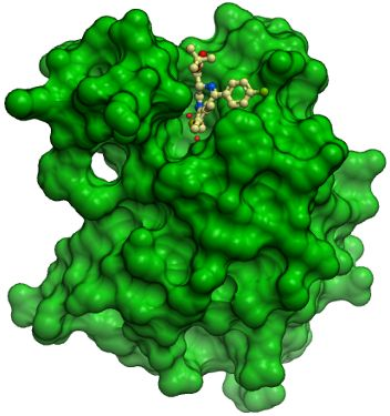
Crystallization: Crystals were grown at 4°C in 300 nl sitting drops from a 1:1 ratio of protein (10.1 mg/ml + 2 mM ligand JQ1/SGCBD01) to reservoir solution containing 0.2 M NaI, 0.1 M BTProp pH 8.5, 20% PEG3350 and 10% ethylene glycol. |
Mass spectrometry characterization: LC- ESI -MS TOF gave a measured mass of 22966 for this construct as predicted from the sequence of this protein. |
Protein concentration: Protein was concentrated to 10 mg/ml using an Amicon 10 kDa cut-off concentrator. |
Crystallisation: Crystals were grown at 20°C in 300 nl sitting drops from a 1:1 ratio of protein to reservoir solution containing 20% PEG-smear (PEG3350 & PEG MME5K), 0.1 M HEPES pH 7.5 |
Data Collection: Crystals were cryo-protected using the well solution supplemented by 25% ethylene glycol and flash frozen in liquid nitrogen.
X-ray source: Diffraction data were collected from a single crystal on a Rigaku FRE with an RAXIS IV detector at a single wavelength of 1.542 Å and the structure was refined to 1.6 Å.
Phasing: The structure was solved by molecular replacement using an ensemble of known bromodomain structures as a starting model. |