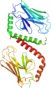
HSPA9
PDB:3N8E
Revision
Revision Type:created
Revised by:created
Revision Date:created
Entry Clone Accession:BC024034
Entry Clone Source:Mammalian Gene Collection
SGC Clone Accession:HSPA9-k015
Tag:N-terminal hexahistidine tag with integrated TEV protease cleavage site: mhhhhhhssgvdlgtenlyfq*sm
Host:E.coli BL21(DE3) R3 pRARE, where R3 denotes a derivative of BL21(DE3) resistant to a strain of T1 bacteriophage (SGC Oxford) and the pRARE plasmid originating from the Rosetta strain (Novagen) supplies tRNAs for rare codons.
Construct
Prelude:Sequence:mhhhhhhssgvdlgtenlyfq*smDVTPLSLGIETLGGVFTKLINRNTTIPTKKSQVFSTAADGQTQVEIKVCQGEREMAGDNKLLGQFTLIGIPPAPRGVPQIEVTFDIDANGIVHVSAKDKGTGREQQIVIQSSGGLSKDDIENMVKNAEKYAEEDRRKKERVEAVNMAEGIIHDTETKME
Vector:pNIC-Bsa4
Growth
Medium:Antibiotics:Procedure:Cells from a glycerol stock were grown in 20 ml TB supplemented with 8 g/l glycerol, 100 µg/ml kanamycin and 34 µg/ml chloramphenicol at 30 °C overnight. The overnight culture (15 ml) was used to inoculate 0.75 l TB supplemented with 8 g/l glycerol and 50 µg/ml kanamycin. The culture was grown at 37 °C until OD600 reached ~1-2. The culture was down-tempered to 18 °C over a period of 60 minutes before target expression was induced by addition of 0.5 mM IPTG. Expression was allowed to continue overnight and cells were harvested the following morning by centrifugation (4,400 x
g, 10 min, 4 °C). The resulting cell pellet (17.1 g wet cell weight) was resuspended in lysis buffer (1.5 ml/g cell pellet), supplemented with 2000 U Benzonase (Merck) and one tablet of Complete EDTA-free protease inhibitor (Roche Applied Science). The cell suspension was stored at -80 °C.
Purification
ProcedureColumnsIMAC: Ni-charged 1 ml HiTrap Chelating HP (GE Healthcare)Gel filtration column: HiLoad 16/60 Superdex 75 Prep Grade (GE Healthcare)
Procedure
IMAC columns were equilibrated with IMAC wash1 buffer, and gel filtration columns were equilibrated with GF buffer. Purification of the protein was performed on an ÄKTA prime system (GE Healthcare). The filtered lysate was loaded onto the Ni-charged HiTrap Chelating column and washed with IMAC wash1 buffer followed by IMAC wash2 buffer. Bound protein was eluted from the IMAC column with IMAC elution buffer and loaded onto the gel filtration column. Fractions containing the target protein were identified by SDS-PAGE and pooled. The protein concentration was 18.26 mg/ml in a volume of 0.75 ml.
Extraction
ProcedureThe cell suspension was quickly thawed in water. Cells were disrupted by sonication (Vibra-Cell, Sonics) at 80% amplitude for 3 min effective time (pulsed 4s on, 4s off) and cell debris was removed by centrifugation (49,000 x
g, 20 min, 4 ºC). The supernatant was decanted and filtered through a 0.45 µm flask filter.
Concentration:LigandMassSpec:Crystallization:Crystals were obtained by the sitting drop vapour diffusion method in a 96-well plate. 0.2 µl of the protein solution was mixed with 0.1 µl of well solution consisting of 0.2 M sodium chloride, 20% PEG 8000, and 0.1 M citric acid, pH 4.2 and 0.1 M di-sodium hydrogen phosphate. The plate was incubated at 20 ºC and crystals appeared within 14 days. The crystals were flash frozen in liquid nitrogen.
NMR Spectroscopy:Data Collection:Diffraction data to 2.8 Å resolution was collected at BESSY (BL14-1).
Data Processing:The structure was solved by molecular replacement using the peptide-binding domain of heat shock 70 kDa protein F, mitochondrial precursor, from Caenorhabditis elegans (PDB:3DQG) as a template. The space group was P6222 with cell dimensions a=118.68 Å, b=118.68 Å, c=159.87 Å, α=90.00, β=90.00, γ=120.00. Two monomers were located in the asymmetric unit. Refmac and autoBUSTER were used for refinement and Coot for model building. Data in the interval 34.94-2.8 Å resolution was used and at the end of the refinement the R values were: R=20.63% and Rfree=24.23%. Coordinates for the crystal structure were deposited in the Protein Data Bank, accession code 3N8E.