GI number: gi|281604136 |
Vector: pNIC28-Bsa4. Details [PDF ]; Sequence [ FASTA ] or [ GenBank ] |
Amplified construct sequence:
CATATGCACCATCATCATCATCATTC
TTCTGGTGTAGATCTGGGTACCGAGA
ACCTGTACTTCCAATCCATGGACCAC
TACAAAGCCAAGGCCACGCGGCACA
TCTTCCTCATCAGGCATTCCCAGTAC
CACGTGGATGGCTCCCTGGAGAAGG
ACCGCACTCTGACCCCGCTGGGTCG
GGAGCAGGCTGAACTCACTGGGCTC
CGCCTGGCAAGCTTGGGGTTGAAGT
TTAATAAAATCGTCCATTCGTCTATG
ACGCGCGCCATAGAGACCACCGAT
ATCATCAGCCGGCACCTGCCAGGCG
TCTGCAAAGTCAGCACAGATCTGCT
GCGGGAAGGCGCCCCCATCGAGCC
AGACCCGCCCGTGTCTCATTGGAA
GCCGGAAGCTGTGCAGTATTACGAA
GACGGAGCCCGGATCGAGGCCGCC
TTCCGGAACTACATCCACCGCGCA
GATGCCAGGCAGGAGGAGGACAGT
TACGAGATCTTCATCTGTCACGCCA
ACGTCATCCGCTACATCGTGTGCAG
AGCACTGCAGTTTCCTCCTGAAGGC
TGGCTCCGGCTCTCCCTCAATAATG
GCAGCATCACCCACCTGGTGATCC
GACCCAACGGCCGAGTTGCGCTCA
GGACCCTCGGGGACACGGGGTTCA
TGCCTCCCGACAAGATCACTCGAT
CCTGACAGTAAAGGTGGATACGGA
TCCGAA
|
Final protein sequence (Tag sequence in lowercase):
mhhhhhhssgvdlgtenlyfq^smDHYKAKATR
HIFLIRHSQYHVDGSLEKDRTLTPLGR
EQAELTGLRLASLGLKFNKIVHSSMT
RAIETTDIISRHLPGVCKVSTDLLREG
APIEPDPPVSHWKPEAVQYYEDGAR
IEAAFRNYIHRADARQEEDSYEIFICH
ANVIRYIVCRALQFPPEGWLRLSLNN
GSITHLVIRPNGRVALRTLGDTGFMPP
DKITRS
^ TEV cleave site |
Tags and additions: Cleavable N-terminal His6 tag. |
Host: BL21 (DE3)R3-pRARE2 (Phage resistant strain).
|
Growth medium, induction protocol: 10 ml from a 50 ml overnight culture containing 50 µg/ml kanamycin and 34 µg/ml chloramphenicol were used to inoculate each of two 1 liter cultures of LB containing 50 µg/ml kanamycin and 34 µg/ml chloramphenicol. Cultures were grown at 37°C until the OD600 reached ~0.6 then the temperature was adjusted to 18°C. Expression was induced overnight using 0.5 mM IPTG at an OD600 of 0.8. The cells were collected by centrifugation and the pellet re-suspended in binding buffer and frozen.
Lysis buffer: 50 mM HEPES pH 7.5; 500 mM NaCl; 10 mM Imidazole, 5% Glycerol.
Extraction buffer, extraction method: Frozen pellets were thawed and fresh 0.5 mM TCEP, 50ml of protease inhibitor cocktail SET VII added to the lysate. Cells were lysed by homogenization. The lysate was centrifuged at 24,000 rpm for 60 minutes and the supernatant collected for purification. |
Column 1: DAE-52, 10 g suspension in 100 ml of 2.5 M NaCl, in a 2 x 25 cm column washed with binding buffer. |
Column 1 Buffer: 50 mM HEPES pH 7.5, 500 mM NaCl, 5% glycerol, 0.5mM TCEP. |
Column 1 Procedure: The supernatant was loaded by gravity flow on the DAE-52 column. The column was then washed with 2 x 50 ml binding buffer at gravity flow. |
Column 2: Ni-affinity. Ni-sepharose (Amersham), 5 ml of 50% slurry in 1.5 x 10 cm column, washed with binding buffer. |
Column 2 Buffers:
Binding buffer: 50 mM HEPES pH 7.5, 500 mM NaCl, 5 mM imidazole, 5% glycerol, 0.5mM TCEP.
Wash buffer: 50 mM HEPES pH 7.5, 500 mM NaCl, 30 mM Imidazole, 5% glycerol, 0.5mM TCEP.
Elution buffer: 50 mM HEPES pH 7.5, 500 mM NaCl, 5% glycerol, 50 to 250 mM Imidazole, 0.5mM TCEP (step elution).
|
Column 2 Procedure: The Ni-sepharose column was serially connected to the DAE-52 column and was loaded by gravity flow. The column was then washed with 30 ml wash buffer at gravity flow. The protein was eluted by gravity flow by applying 5-ml portions of elution buffer with increasing concentration of imidazole (50 mM, 100 mM, 150 mM, 200 and 250 mM); fractions were collected until essentially all protein was eluted. |
Enzymatic treatment: The N-terminal His tag was cleaved by treatment with TEV protease, overnight. |
Column 3: Size Exclusion Chromatography. Superdex S200 16/60 HiLoad. |
Column 3 Buffer: 50 mM Tris.HCl, pH 7.5; 300 mM NaCl, 0.5 mM TCEP. |
Column 3 Procedure: The protein was concentrated and applied to an S200 16/60 HiLoad gel filtration column equilibrated in 50 mM Tris.HCl, pH 7.5; 300 mM NaCl, 0.5 mM TCEP using an ÄKTA express system. |
Mass spectrometry characterization: LC- ESI -MS TOF gave a measured mass of 22966 for this construct as predicted from the sequence of this protein. |
Protein concentration: Protein was concentrated to 10 mg/ml using an Amicon 10 kDa cut-off concentrator. |
Crystallisation: Prior to crystallization, 20mM Na/K phosphate was added into protein solution. Crystals were grown at 20°C in 300 nl sitting drops from a 1:1 ratio of protein to reservoir solution containing 15% PEG-smear (PEG3350 & PEG MME5K), 0.1 M HEPES pH 7.5 |
Data Collection: Crystals were cryo-protected using the well solution supplemented by 20% ethylene glycol and flash frozen in liquid nitrogen.
X-ray source: Diffraction data were collected from a single crystal on a Rigaku FRE with an RAXIS IV detector at a single wavelength of 1.542 Å. The final structure was refined to 1.90 Å.
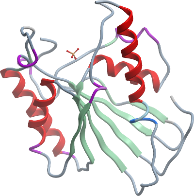
Phasing: The structure was solved by molecular replacement using our previous apo-form PGAM5 as a starting model. |