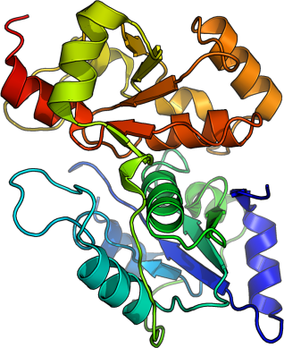
MPST
PDB:3OLH
Revision
Revision Type:created
Revised by:created
Revision Date:created
Entry Clone Accession:BC016737
Entry Clone Source:MGC
SGC Clone Accession:MPSTA-s001
Tag:N-terminal hexahistidine tag: mhhhhhhssgvdlgtenlyfqsm
Host:Escherichia coli BL21(DE3) R3 pRARE
Construct
Prelude:Sequence:mhhhhhhssgvdlgtenlyfqsm*VSAQWVAEALRAPRAGQPLQLLDASWYLPKLGRDARREFEERHIPGAAFFDIDQCSDRTSPYDHMLPGAEHFAEYAGRLGVGAATHVVIYDASDQGLYSAPRVWWMFRAFGHHAVSLLDGGLRHWLRQNLPLSSGKSQPAPAEFRAQLDPAFIKTYEDIKENLESRRFQVVDSRATGRFRGTEPEPRDGIEPGHIPGTVNIPFTDFLSQEGLEKSPEEIRHLFQEKKVDLSKPLVATCGSGVTACHVALGAYLCGKPDVPIYDGSWVEWYMRARPEDVI
Vector:pNIC-Bsa4
Growth
Medium:Antibiotics:Procedure:Fresh overnight cultures of
E. coli strain BL21(DE3) R3 pRARE cells (including 100 µg/ml kanamycin and 34 µg/ml chloramphenicol) transformed with MPST expression construct were used to inoculate 4.5 l TB supplemented with 8 g/l glycerol, 50 µg/ml kanamycin and anti-foam (Dow Corning) in three 2-liter flasks. Cells were grown in a large scale expression system (Harbinger Biotechnology and Engineering) at 37°C until the OD
600 reached ~2. The culture was down-tempered to 18°C for 1 h. Expression of MPST was induced by adding 0.5 mM IPTG and growth continued over night at 18°C. Cells were harvested by centrifugation at 4400 x
g for 10 min. The pellet was resuspended in lysis buffer supplemented with Complete EDTA-free Protease Inhibitor (Roche Biosciences) and benzonase. Suspended cells were stored at -80°C until further use.
Purification
ProcedureColumnsIMAC: Ni-charged 2 x 1 ml HiTrap Chelating HP (GE Healthcare) in series
Gel filtration column: HiLoad 16/60 Superdex 200 (GE Healthcare)
PurificationPurification of the protein was performed on an ÄKTAxpress system (GE Healthcare). Prior to purification, IMAC column was equilibrated with IMAC wash1 buffer and gel filtration column with gel filtration buffer. The filtered lysate was loaded onto the IMAC column, and therafter washed with IMAC wash1 buffer followed by IMAC wash2 buffer. Bound protein was eluted from the IMAC column with IMAC elution buffer and loaded onto the gel filtration column. Eluting fractions were analyzed by SDS-PAGE and target protein was pooled. Fresh TCEP was added to a final concentration of 2 mM. Purified protein was concentrated using an Amicon Ultra-15 centrifugal filter device (Millipore, 10,000 MWCO) to 26.4 mg/ml.
Extraction
ProcedureThe cell suspension was quickly thawed in warm water. Cells were disrupted by sonication (Vibra-Cell, Sonics) at 80% amplitude for 3 min effective time (pulsed 4s on, 4s off) and cell debris was removed by centrifugation (49000 x
g, 20 min, 4 ºC). The supernatant was decanted and filtered through a 0.45 µm flask filter.
Concentration:LigandMassSpec:Crystallization:Crystals were obtained by the sitting drop vapour diffusion method in a 96-well plate. 0.2 µl protein solution (25.5 mg/ml) including 20mM sodium pyruvate was mixed with 0.1 µl of well solution consisting of 1.5M ammonium sulfate, 0.1M sodium acetate pH 3.9. The plate was incubated at 4 ºC. Crystals appeared after 5 days and continued to grew for one more week wherafter they were quickly transferred to a cryo solution consisting of 1.7M ammonium sulfate, 0.1M sodium acetate pH 4.6, 27% Glycerol, 0.2M sodium chloride and flash frozen in liquid nitrogen.
NMR Spectroscopy:Data Collection:Diffraction data to 2.50 Å resolution was collected at BESSY beamline BL14-2.
Data Processing:Data were indexed and integrated in space group R32 using XDS software. The structure was solved by molecular replacement using the structure of Bos taurus Rhodanese (pdb: 1boi) as model template. Balbes was used to solve the structure. The asymmetric unit contained one protein monomer. The cell dimensions are a = 171.3Å, b = 171.3Å, c = 75.8Å. Buster was used for refinement and Coot for manual model building. TLS refinement was used, four groups as suggested by TLSMD-server (
http://skuld.bmsc.washington.edu/~tlsmd/). Data in the interval 33.7-2.50 Å resolution were used and refined to R = 22.61% and Rfree = 27.01%. Coordinates for the crystal structure were deposited in the Protein Data Bank, with accession code 3olh.