Entry Clone Source: Site-directed mutagenesis |
Entry Clone Accession: n/a |
SGC Construct ID: ACVR1A-c096 |
GenBank GI number: gi|4501895 |
Vector: pFB-LIC-Bse. Details [ PDF ]; Sequence [ FASTA ] or [ GenBank ] |
Entry Clone Source: Site-directed mutagenesis |
Amplified construct sequence:
CCATGGGCCACCATCATCATCATCATTCTT
CTGGTGTAGATCTGGGTACCGAGAACCTGT
ACTTCCAATCCATGCAAAGAACAGTGGCTC
GCGATATTACACTGTTGGAGTGTGTCGGGA
AAGGCAGGTATGGTGAGGTGTGGAGGGGCA
GCTGGCAAGGGGAAAATGTTGCCGTGAAGA
TCTTCTCCTCCCGTGATGAGAAGTCATGGT
TCAGGGAAACGGAATTGTACAACACTGTGA
TGCTGAGGCATGAAAATATCTTAGGTTTCA
TTGCTTCAGACATGACATCAAGACACTCCA
GTACCCAGCTGTGGTTAATTACACATTATC
ATGAAATGGGATCGTTGTACGACTATCTTC
AGCTTACTACTCTGGATACAGTTAGCTGCC
TTCGAATAGTGCTGTCCATAGCTAGTGGTC
TTGCACATTTGCACATAGAGATATTTGGGA
CCCAAGGGAAACCAGCCATTGCCCATCGAG
ATTTAAAGAGCAAAAATATTCTGGTTAAGA
AGAATGGACAGTGTTGCATAGCAGATTTGG
GCCTGGCAGTCATGCATTCCCAGAGCACCA
ATCAGCTTGATGTGGGGAACAATCCCCGTG
TGGGCACCAAGCGCTACATGGCCCCCGAAG
TTCTAGATGAAACCATCCAGGTGGATTGTT
TCGATTCTTATAAAAGGGTCGATATTTGGG
CCTTTGGACTTGTTTTGTGGGAAGTGGCCA
GGCGGATGGTGAGCAATGGTATAGTGGAGG
ATTACAAGCCACCGTTCTACGATGTGGTTC
CCAATGACCCAAGTTTTGAAGATATGAGGA
AGGTAGTCTGTGTGGATCAACAAAGGCCAA
ACATACCCAACAGATGGTTCTCAGACCCGA
CATTAACCTCTCTGGCCAAGCTAATGAAAG
AATGCTGGTATCAAAATCCATCCGCAAGAC
TCACAGCACTGCGTATCAAAAAGACTTTGA
CCAAAATTGATTGACAGTAAAGGTGGATAC
GGATCCGAATTCGAGCTCCGTCGACAAGCT
T |
Final protein sequence (tag sequence in lowercase):
mghhhhhhssgvdlgtenlyfqsMQRTVAR
DITLLECVGKGRYGEVWRGSWQGENVAVKI
FSSRDEKSWFRETELYNTVMLRHENILGFI
ASDMTSRHSSTQLWLITHYHEMGSLYDYLQ
LTTLDTVSCLRIVLSIASGLAHLHIEIFGT
QGKPAIAHRDLKSKNILVKKNGQCCIADLG
LAVMHSQSTNQLDVGNNPRVGTKRYMAPEV
LDETIQVDCFDSYKRVDIWAFGLVLWEVAR
RMVSNGIVEDYKPPFYDVVPNDPSFEDMRK
VVCVDQQRPNIPNRWFSDPTLTSLAKLMKE
CWYQNPSARLTALRIKKTLTKID
Engineered Q207D mutation in bold and underlined. |
Tags and additions: mghhhhhhssgvdlgtenlyfq*sM. cleavable N-terminal hexahistidine tag. |
Host: SF9 Spodoptera frugiperda Insect cells
|
Growth medium, induction protocol: Sf9 cells at a density of 2x10⁶/ml were infected with recombinant ACVR1 baculovirus (virus stock P3; 1ml of virus stock/100 ml of cell culture). Cells were shaken at 110 rpm at 27°C in an Innova shaker. After 48 hours post-infection the cultures were harvested by centrifugation for 20min at 6000rpm. Cell pellets from each 1L flask were resuspended in 15 ml binding buffer.
Binding buffer: 50 mM Hepes, pH 7.5; 500 mM NaCl; 5% Glycerol; 5 mM imidazole.
Extraction buffer, extraction method: The frozen cells were thawed and the volume increased to 200 ml with binding buffer. The cells were lysed using an Emulsiflex C5 homogeniser. The cell lysate was spun down by centrifugation at 21K rpm and 4°C for 1 h. The supernatant was recovered for purification. |
Column 1: Anion-exchange for Nucleic acid removal with DEAE cellulose (DE52, Whatmann) 10g of resin was suspended in 50 ml 1 M NaCl, and then applied onto a 2.5 x 20 cm column. The resin was then equilibrated with 50 ml binding buffer prior to loading the sample. |
Buffers:
Binding buffer: 50 mM Hepes, pH 7.5; 500 mM NaCl; 5% Glycerol; 5 mM imidazole, 0.1mM TCEP
Wash buffer: 50 mM Hepes, pH 7.5; 500 mM NaCl; 5% Glycerol; 25 mM imidazole, 0.1mM TCEP |
Column 1 Procedure: The supernatant was first applied onto the column by gravity flow, which was followed by a wash with 50 ml wash buffer. The column flow-through and wash was directly applied onto a Ni-sepharose column. |
Column 2: Ni-Affinity Chromatography. 6 ml of 50% Ni-sepharose slurry was applied onto a 1.5 x 10 cm column. The column was equilibrated with binding buffer (25ml). |
Column 2 Buffers:
Binding buffer: 50 mM Hepes, pH 7.5; 500 mM NaCl; 5% Glycerol; 5 mM imidazole, 0.1mM TCEP
Wash buffer: 50 mM Hepes, pH 7.5; 500 mM NaCl; 5% Glycerol; 25 mM imidazole, 0.1mM TCEP
Elution buffer: 50 mM HEPES, pH 7.5; 500 mM NaCl; 5% Glycerol; 50 to 250 mM imidazole, 0.1mM TCEP |
Column 2 Procedure: The flow-through from column 1 (DE52) was applied by gravity flow onto the Ni-sepharose column. The bound protein was eluted by applying a step gradient of imidazole - using 10 ml portions of elution buffer with increasing concentration of imidazole (50 mM, 100 mM, 150 mM, 250 mM). |
Enzymatic treatment:0.1mg of TEV protease was added to the Ni-eluted protein to remove the tag. |
Column 3: Size Exclusion Chromatography - S200 HiLoad 16/60 Superdex run on ÄKTA-Express |
Column 3 Buffers: Gel Filtration buffer: 300 mM NaCl, 50 mM Hepes pH 7.5, 0.5mM TCEP |
Column 3 Procedure: Prior to applying the protein, the S200 16/60 column was washed and equilibrated with gel filtration buffer. The protein was concentrated to 5 ml using an Amicon Ultra-15 filter with a 10 kDa cut-off. The concentrated protein was directly applied onto the equilibrated S200 16/60 column, and run at a flow-rate of 1 ml/min. The protein was eluted at 90-108 ml. Fractions containing the protein were pooled together. |
Column 4: Ni-Affinity Chromatography. 0.75 ml of 50 % Ni-sepharose slurry was applied onto a 1.5 x 10 cm column. The column was equilibrated with binding buffer (15ml). |
Column 4 Buffers:
Binding buffer: 50 mM Hepes, pH 7.5; 500 mM NaCl; 5% Glycerol; 5 mM imidazole, 0.1mM TCEP
Wash buffer: 50 mM Hepes, pH 7.5; 500 mM NaCl; 5% Glycerol; 25 mM imidazole, 0.1mM TCEP
Elution buffer: 50 mM HEPES, pH 7.5; 500 mM NaCl; 5% Glycerol; 250 mM imidazole, 0.1mM TCEP |
Column 4 Procedure: The cleaved protein was passed through the column followed by 3ml binding buffer. It was then washed with 8ml wash buffer. Anything remaining bound to the column was eluted with 15ml elution buffer. |
Mass spec characterization: The purified protein was homogeneous and had an experimental mass of 34494.5 (after TEV cleavage), closely matching the expected mass 34492.7. Mass was determined by LC-MS, using an Agilent LC/MSD TOF system with reversed-phase HPLC coupled to electrospray ionisation and an orthogonal time-of-flight mass analyser. Proteins were desalted prior to mass spectrometry by rapid elution off a C3 column with a gradient of 5-95% isopropanol in water with 0.1% formic acid. |
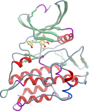
Crystallisation: Protein was buffered in 50 mM HEPES pH 7.5, 300 mM NaCl, 10 mM DTT and 10mM L-arginine, 10 mM L-glutamate. The protein was concentrated to 10 mg/ml (calculated using an extinction co-efficient of 58900) in the presence of the inhibitor K00507a (1 mM end concentration). Crystals were grown at 20°C in 150 nl sitting drops mixing 50 nl protein solution with 100 nl of a reservoir solution containing 0.2M Na/KPO4, 20% PEG 3350, 10% ethylene glycol. On mounting crystals were cryoprotected with mother liquor plus 20% ethylene glycol before transfer to liquid nitrogen. |
Data Collection:
Resolution: 2.0 Å resolution
X-ray source: Diamond Light Source, station I03 |