|
Entry Clone Source: MGC
|
|
Entry Clone Accession: BC028915
|
|
SGC Construct ID: CBLCA-c012
|
|
GenBank GI number: gi|195927027
|
|
Vector: pNIC-CTHF. Details [ PDF ]; Sequence [ FASTA ] or [ GenBank ]
|
|
Amplified construct sequence:
TTAAGAAGGAGATATACTATGGGGCGACAG
TGGGAAGAGGCCCGCGCCCTGGGCCGGGCA
GTCAGGATGCTGCAGCGCCTAGAAGAGCAA
TGCGTCGACCCCCGGCTGTCCGTGAGTCCC
CCTTCGCTGCGGGACCTGCTGCCCCGCACA
GCGCAGCTGCTTCGAGAGGTGGCCCATTCT
CGGCGGGAGGCCGGCGGAGGCGGCCCCGGG
GGTCCCGGCGGCTCTGGGGACTTTCTACTC
ATCTACCTGGCCAATCTGGAGGCCAAGAGC
AGGCAGGTGGCCGCGCTGCTGCCTCCCCGG
GGCCGAAGGAGTGCCAACGACGAGCTCTTC
CGGGCGGGCTCCAGACTCAGGCGACAGCTG
GCCAAGCTGGCCATCATCTTCAGCCACATG
CACGCAGAGCTGCACGCACTCTTCCCCGGG
GGAAAGTACTGTGGACACATGTACCAGCTC
ACCAAGGCCCCCGCCCACACCTTCTGGAGG
GAAAGTTGCGGAGCCCGGTGTGTGCTGCCC
TGGGCTGAGTTTGAGTCCCTCCTGGGCACC
TGCCACCCTGTGGAACCAGGCTGCACAGCC
CTGGCCTTGCGCACCACCATTGACCTCACC
TGCAGCGGGCACGTGTCCATCTTCGAGTTC
GACGTCTTCACCAGGCTCTTTCAGCCATGG
CCAACACTCCTCAAGAACTGGCAGCTCCTG
GCAGTCAACCACCCAGGCTACATGGCCTTC
CTCACCTATGATGAGGTCCAAGAGCGTCTG
CAGGCCTGCAGGGACAAGCCAGGCAGTTAC
ATCTTCCGGCCCAGCTGTACTCGCCTGGGG
CAGTGGGCCATCGGCTATGTGAGCTCAGAT
GGCAGCATCCTGCAGACCATCCCTGCCAAC
AAACCCCTGTCCCAGGTGCTCCTGGAGGGA
CAGAAGGACGGCTTCTACCTCTACCCAGAT
GGAAAGACCCACAACCCAGACCTGACTGAG
CTCGGCGCAGAGAACCTCTACTTCCAATC
Note a mutation was identified by DNA sequencing with the codon change from GCG to GAG (shown bold) resulting in the amino acid change A64E
|
|
Final protein sequence (tag sequence in lowercase):
MGRQWEEARALGRAVRMLQRLEEQCVDPRL
SVSPPSLRDLLPRTAQLLREVAHSRREAGG
GGPGGPGGSGDFLLIYLANLEAKSRQVAAL
LPPRGRRSANDELFRAGSRLRRQLAKLAII
FSHMHAELHALFPGGKYCGHMYQLTKAPAH
TFWRESCGARCVLPWAEFESLLGTCHPVEP
GCTALALRTTIDLTCSGHVSIFEFDVFTRL
FQPWPTLLKNWQLLAVNHPGYMAFLTYDEV
QERLQACRDKPGSYIFRPSCTRLGQWAIGY
VSSDGSILQTIPANKPLSQVLLEGQKDGFY
LYPDGKTHNPDLTELGAENLYFq*shhhhh
hdykddddk
^ TEV cleave site
|
|
Tags and additions: C-terminal TEV-cleavable (at *) his-tag with the following sequence: Q*SHHHHHHDYKDDDDK
|
|
Host: BL21 (DE3)R3-pRARE2 (Phage resistant strain)
|
|
Growth medium, induction protocol: 10ml of overnight culture from grown from a glycerol stock and finally added to 1L LB flasks with 50mg/ml of Kanamycin and 34mg/ml of chloramphenicol (total 12L). The cells were cultured at 37°C until the OD reached 0.8 and then decreased to 18°C. IPTG was added at 0.2mM (final concentration) for induction at 18°C overnight.
Binding buffer: 500 mM NaCl, 5% Glycerol, 50 mM HEPES pH 7.5, 30 mM Imidazole. Complete Protease Inhibitor Cocktail Tablets (Roche) were added (one tablet/50ml buffer).
Extraction buffer, extraction method: The cells were harvested by centrifugation at 4,000 g for 10 min. The pellet from 1 L culture was resuspended in 25 ml of extraction buffer. The sample was lysed by sonication and then centrifuged at 37505 g for 30 minutes. The supernatant was kept for further purification.
|
|
Column 1: Ni-sepharose
|
|
Column 1 Buffers:
Binding buffer: 500 mM NaCl, 5% Glycerol, 50 mM HEPES pH 7.5, 30 mM Imidazole.
Washing Buffer: 500 mM NaCl, 5% Glycerol, 50 mM HEPES pH 7.5, 30 mM Imidazole.
Elution Buffer: I: 500 mM NaCl, 5% Glycerol, 50 mM HEPES pH 7.5, 60 mM Imidazole.
Elution Buffer II: 500 mM NaCl, 5% Glycerol, 50 mM HEPES pH 7.5, 125 mM Imidazole.
Elution Buffer III: 500 mM NaCl, 5% Glycerol, 50 mM HEPES pH 7.5, 250 mM Imidazole.
|
|
Column 1 Procedure: The column was packed with 6ml of Ni-sepharose slurry and equilibrated with 15ml of binding buffer. The supernatant was loaded onto the column and the flow through was collected. The column was washed sequentially with 10ml of washing buffer followed by a second 20 ml wash fraction. The protein was then eluted with a step gradient of 10ml fractions of elution buffer I, II and III, respectively.
|
|
Enzymatic treatment: 400µl of TEV protease (6mg/ml) were added into the sample from Ni-purification (fractions wash II, elute I, II and III). The sample was incubated at 4°C overnight.
|
|
Column 2: Superdex 200 Hiload 16/60
|
|
Column 2 Buffers: 500 mM NaCl, 5% Glycerol, 50 mM HEPES pH 7.5, 0.5 mM TCEP.
|
|
Column 2 Procedure: After TEV cleavage, the sample was concentrated to 3ml before loading onto an ÄKTA Purifier. Gel filtration was run at 4°C. Elution fractions were analyzed by SDS-PAGE and the most purified fractions were collected.
|
|
Column 3: Ni-sepharose
|
|
Column 3 Buffers: 500 mM NaCl, 5% Glycerol, 50 mM HEPES pH 7.5, 0.5 mM TCEP.
|
|
Column 3 Procedure: The cleaved sample was loaded onto the column (packed from 0.5ml of Ni-sepharose slurry). The flow through was collected and the column was then washed with 5ml of the buffer (also collected).
|
|
Protein concentration: Protein was concentrated to 10 mg/ml using an Amicon 10 kDa cut-off concentrator.
|
|
Mass spectrometry characterization: Masses of purified proteins were confirmed by LC-MS, using an Agilent LC/MSD TOF system with reversed-phase HPLC coupled to electrospray ionisation and an orthogonal time-of-flight mass analyser. Proteins were desalted prior to mass spectrometry by rapid elution off a C3 column with a gradient of 5-95% isopropanol in water with 0.1% formic acid. A mass of 35952.2 was observed suggesting that the N-terminal methionine was lost.
|
|
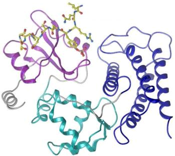
Crystallisation: Crystals were obtained at 4°C in 150 nl sitting drops set up at a ratio of 1:1 with mother liquor and 12.3mg/ml protein with 1mM of EGFR peptide (pY1069). The mother liquor consisted of 0.1 M HEPES pH 7.5 and 20% (v/v) jeffamine M-600. On mounting crystals were cryo-protected with an additional 10% PEG400.
|
|
Structure Determination:
Resolution: 2.5 Å resolution.
X-ray source: Diamond Light Source, station I02, using monochromatic radiation at wavelength 0.9795 Å.
|