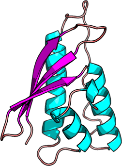
NISCH
PDB:3P0C
Revision
Revision Type:created
Revised by:created
Revision Date:created
Entry Clone Accession:BC038102
Entry Clone Source:Mammalian Gene Collection
SGC Clone Accession:NISCHA-s002
Tag:N-terminal hexahistidine tag with integrated TEV protease cleavage site: mhhhhhhssgvdlgtenlyfq*sm
Host:E.coli BL21(DE3) R3 pRARE, where R3 denotes a derivative of BL21(DE3) resistant to a strain of T1 bacteriophage (SGC Oxford) and the pRARE plasmid originating from the Rosetta strain (Novagen) supplies tRNAs for rare codons.
Construct
Prelude:
Sequence:mhhhhhhssgvdlgtenlyfqsmEARVVGSELVDTYTVYIIQVTDGSHEWTVKHRYSDFHDLHEKLVAERKIDKNLLPPKKIIGKNSRSLVEKREKDLEVYLQKLLAAFPGVTPRVLAHFLHFHFYEING
Vector:pNIC-Bsa4
Growth
Medium:
Antibiotics:
Procedure:Cells from a glycerol stock were grown in 20 ml TB supplemented with 8 g/l glycerol, 100 µg/ml kanamycin and 34 µg/ml chloramphenicol at 30 °C overnight. The overnight culture (10 ml) was used to inoculate 750ml TB supplemented with 8 g/l glycerol, 50 µg/ml kanamycin and approximately 200 µl 204 Antifoam A6426 (Sigma). The culture was grown in a LEX bioreactor system (Harbinger Biotechnology) at 37 °C until OD600 reached ~2. The culture was down-tempered to 18 °C over a period of 1 hour before target expression was induced by addition of 0.5 mM IPTG. Expression was allowed to continue overnight and cells were harvested the following morning by centrifugation (4,400 x g, 10 min, 4 °C). The resulting cell pellet (12 g wet cell weight) was resuspended in lysis buffer (1.5 ml/g cell pellet), supplemented with 2000 U Benzonase (Merck) and one tablet of Complete EDTA-free protease inhibitor (Roche Applied Science). The cell suspension was stored at -80 °C.
Purification
Procedure
Columns
IMAC: Ni-charged 1 ml HiTrap Chelating HP (GE Healthcare)
Gel filtration column: HiLoad 16/60 Superdex 75 Prep Grade (GE Healthcare)
Procedure
IMAC columns were equilibrated with IMAC wash1 buffer, and gel filtration columns were equilibrated with GF buffer. Purification of the protein was performed on an ÄKTAxpress system (GE Healthcare). The filtered lysate was loaded onto the Ni-charged HiTrap Chelating column and washed with IMAC wash1 buffer followed by IMAC wash2 buffer. Bound protein was eluted from the IMAC column with IMAC elution buffer and automatically loaded onto the gel filtration column. Fractions containing the target protein were identified by SDS-PAGE, pooled, and fresh TCEP was added to a final concentration of 2 mM. The protein was concentrated using an Amicon Ultra-15 centrifugal filter device (10,000 NMWL; Millipore) to 17.2 mg/ml in a volume of 1 ml. The identity of the protein was confirmed by mass spectrometry.
Extraction
Procedure
The cell suspension was quickly thawed in water. Cells were disrupted by sonication (Vibra-Cell, Sonics) at 80% amplitude for 3 min effective time (pulsed 4s on, 4s off) and cell debris was removed by centrifugation (49,000 x g, 20 min, 4 ºC). The supernatant was decanted and filtered through a 0.45 µm flask filter.
Concentration:
Ligand
MassSpec:
Crystallization:Crystals were obtained by the sitting drop vapour diffusion method in a 96-well plate. 0.2 µl protein solution (17.2mg/ml) was mixed with 0.1 µl of well solution consisting of PEG monomethyl ether 2000 11%-20%, Tris 0.1M pH=9.0, trimetylamine n-oxide 0.3-0.2M, benzene 1,2,4-trisphosphate 1mM .The plate was incubated at 4 ºC. Crystals appeared after 5 days and continued to grow for four more weeks. They were quickly transferred to a cryo solution consisting 1,2-Propanediol 20%, PEG monomethyl ether 2000 20%, NaCl 0.3M, Tris 0.1M pH 9, trimetylamine n-oxide 0.2M, benzene 1,2,4-trisphosphate 1mM and flash frozen in liquid nitrogen. The crystal that was used for the SIRAS experiment was soaked with 1mM HgCl2 in the crystallization buffer over night
NMR Spectroscopy:
Data Collection:Data were collected at the DIAMOND Beamline I03 on the 2010-07-08. The peak wavelength for the SIRAS-experiment (Hg soaked crystal) was 1.00730 Å. The crystal diffracted to 2.7 Å. A second dataset with an unsoaked crystal was collected at 0.97920 Å. This crystal diffracted to 2.27 Å
Data Processing:Indexing and intensity integration was performed by MOSFLM. For data scaling SCALA was used (Spacegroup: P212121 Unit Cell: a = 57.88Å, b =64.78Å, c = 80.68Å, α = β = γ = 90°). The structure was solved by Auto-Rickshaw using the SIRAS procedure. Phase extension and structure refinement was done by PHENIX using the dataset of the unsoaked crystal. Two monomers were found in the asymmetric unit, which possibly would form a dimer in solution.
Resolution: 2.27Å
Rmerge = 0.09
R = 0.176
Rfree = 0.210.