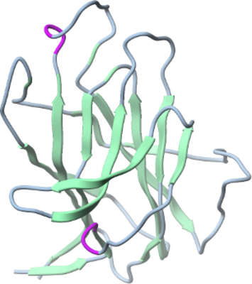
EPHB3
PDB:3P1I
Revision
Revision Type:created
Revised by:created
Revision Date:created
Entry Clone Accession:AT75-A1
Entry Clone Source:Mammalian Gene Collection
SGC Clone Accession:ephb3.0039.0211.173C08 (SDC173C08)
Tag:AAPEHHHHHHDYDIPTTENLYFQGAMD
Host:
Construct
Prelude:
Sequence:EETLMDTKWVTSELAWTSHPESGWEEVSGYDEAMNPIRTYQVCNVRESSQNNWLRTGFIWRRDVQRVYVELKFTVRDCNSIPNIPGSCKETFNLFYYEADSDVASASSPFWMENPYVKVDTIAPDESFSRLDAGRVNTKVRSFGPLSKAGFYLAFQDQGACMSLISVRAFYKK
Vector:pFHMSP-LIC-C
Growth
Medium:Serum Free Medium (HyClone SFX-Insect, SH3027802)
Antibiotics:
Procedure:Sf9 cells were grown in Serum Free Medium (HyClone SFX-Insect, SH3027802) at density of 3.5 million cells per milliliter of media and with viability not less then 97% were infected with 10 mL of P3 viral stock for each 1 L of cell culture. Cell culture medium was collected after 3-4 days of incubation on a shaker at 100 rpm and 27 °C when cells viability dropped to 45-65%.
Purification
Procedure
The cultured medium was centrifuged at 14,000 xg for 15 minutes, and the pH of the supernatant was adjusted to 7.5 at room temperature by adding Titrating Buffer. PMSF (Bioshop, PMS 123.50) and benzamidine (Sigma, B6506-100G) were added to final concentrations of 1 and 2 mM, respectively. A 3.2 L volume of medium was mixed with 30 ml pre-equilibrated NiNTA Superflow beads (Qiagen, 30450) and stirred (Talboys, 134-2) on ice for 1 hour. The resin was transferred to a 100 ml gravity column, washed with 300 ml of Wash Buffer and the protein was eluted with 30-40 ml of Elution Buffer. A second round of NiNTA batch absorption may have been performed for increased yield. The eluate was dialyzed against 50-100 X volume of buffer A overnight at 4 °C. Anion exchange chromatography has been done on HitrapQ HP column (Amersham-Pharmacia, 17-1154-01) using AKTA purifier 100 system. Elution gradient between buffer A and buffer B has been employed. Collected protein fractions were concentrated using 15 mL concentrators with an appropriate molecular weight cut-off (Amicon Ultra-15 10,000 MWCO, Millipore, UFC 901024) to a final value of 10 mg/mL. The average yield was about 6 mg per liter of insect cell culture. Coomassie-stained SDS-PAGE showed that the product was pure and mass-spectroscopy by LC/MS (Agilent 1100 Series) showed that the protein had the correct molecular weight with glycosylation at a single site.
Extraction
Procedure
Concentration:
Ligand
MassSpec:MW =23072.64 g/mol
Crystallization:Crystals were grown at 18 °C using the hanging drop method in greased VDX Plates (Hampton Research, HR3-306) by mixing equal volumes of protein (9.6 mg/ml) and Crystallization Buffer (1.6 M NH4SO4, 5% isopropanol, 0.1 M Tris pH 8.0). Prior to dunking and storage in liquid nitrogen, suitable crystals were immersed in paratone/mineral oil (50/50 by volume).
NMR Spectroscopy:
Data Collection:Diffraction data was collected on the CLSI BEAMLINE 08ID-1 and integrated and scaled using the HKL2000 program suite. The structure was solved by molecular replacement techniques using the program BALBES and search model PDB entry 3EPT. Automated model building using ARP/wARP, combined with iterative model building using the graphics program Coot and maximum-likelihood and TLS refinement with the program REFMAC5 led to a model with an Rfactor of 18.4% (Rfree 23.5%) for data between 19.75 to 2.1 Å.
Data Processing: