Entry Clone Source: Synthetic |
Entry Clone Accession: n/a |
SGC Construct ID: MAST1A-c018 |
GenBank GI number: gi|45120119 |
Vector: pNIC28-Bsa4. Details [PDF ]; Sequence [ FASTA ] or [ GenBank ] |
Amplified construct sequence:
ATGCACCATCATCATCATCATTCTTC
TGGTGTAGATCTGGGTACCGAGAACC
TGTACTTCCAATCCATGCGTAGCCCG
ATTACCATTCAGCGTAGCGGCAAAAA
ATATGGTTTTACCCTGCGTGCGATTC
GCGTGTATATGGGCGATACCGATGTT
TATAGCGTTCATCATATTGTTTGGCA
TGTGGAAGAAGGTGGTCCGGCGCAGG
AAGCGGGCCTGTGCGCGGGCGATCTG
ATTACCCATGTGAATGGTGAACCGGT
GCATGGCATGGTTCATCCGGAAGTGG
TTGAACTGATTCTGAAAAGCGGTAAT
AAAGTGGCGGTGACCACCACCCCGTT
TGAAAACACCCAGAGCCTGGTTTGA
|
Final protein sequence (Tag sequence in lowercase):
smRSPITIQRSGKKYGFTLRAIRVYM
GDTDVYSVHHIVWHVEEGGPAQEAGL
CAGDLITHVNGEPVHGMVHPEVVELI
LKSGNKVAVTTTPFENTQSLV
The N-terminal 2 residues, sm, derive from the vector, following TEV protease cleavage of the hexahistidine tag. |
Tags and additions: Cleavable N-terminal His6 tag. |
Host: BL-21(DE3)-R3-Rosetta (A homemade phage resistant version of BL21(DE3) containing the pRARE2 plasmid from Rosetta II (DE3) cells)
|
Growth medium, induction protocol: A number of colonies from the transformation were used to inoculate 1ml of LB media containing 50µg/ml kanamycin and 34µg/ml chloramphenicol, which was placed in a 37°C shaker overnight. The next day glycerol stocks were prepared from this overnight culture. A glycerol stock was used to inoculate 50ml of TB media containing 50µg/ml kanamycin and 34µg/ml chloramphenicol, which was placed in a 37°C shaker overnight. The next day this starter culture was used to inoculate 2x 1L of TB media (18ml starter culture into each) containing 33µg/ml kanamycin. After 3 hours the temperature was reduced to 20°C. After a further 30 minutes the cells were induced by the addition of 0.5 mM IPTC. The expression was continued overnight. Cells were spun at 6000rpm for 15 minutes at 4°C. The cells were resuspended in 30ml of Lysis Buffer with the addition of 0.2 mM PMSF. The resuspended cell pellet was placed in a -80°C.
Lysis Buffer: 50 mM HEPES pH 7.5; 500 mM NaCl; 5% Glycerol; 5 mM Imidazole, pH 7.5; 0.5 mM TCEP.
Extraction buffer, extraction method: The resuspended cell pellet was lysed by sonication. PEI (polyethlyeneimine) was added to a final concentration of 0.25% and the cell debris and preciptated DNA were spun down (18000rpm, JA18 rotor, 40 min). |
Column 1: Ni-NTA (2ml volume in a gravity-flow column). |
Column 1 Buffer:
Binidng Buffer: 50 mM HEPES, pH 7.5; 500 mM NaCl; 5% glycerol; 5 mM Imidazole, pH 7.5; 0.5mM TCEP.
Wash Buffer:50 mM HEPES, pH 7.5; 500 mM NaCl; 5% Glycerol; 25 mM Imidazole, pH 7.5; 0.5 mM TCEP.
Elution Buffer: 50 mM HEPES, pH 7.5; 500 mM NaCl; 5% Glycerol; 250 mM Imidazole, pH 7.5; 0.5 mM TCEP. |
Column 1 Procedure: The clarified cell extract was passed through the column. The column was then washed with Binding Buffer (25ml) and Wash Buffer (25ml). The protein was eluted with 10ml of Elution Buffer. |
Column 2: S200 16/60 Gel Filtration. |
Column 2 Buffers: 25 mM HEPES, pH 7.5; 150 mM NaCl; 0.5 mM TCEP. |
Column 2 Procedure: The wash 2 and elution fractions from column 1 were combined and concentrated to 4ml volume in a 3kDa MW cutoff spin concentrator.The concentrated sample was injected onto an S200 16/60 column (pre-equilibrated in GF Buffer) at 1.0ml/min. 1.75ml fractions were collected. |
TEV protease digestion: Gel filtrations containing MAST1A were combined. TEV protease was added and the sample left at 4°C over night. |
Rebinding of impurities to Ni-NTA: The protein was passed through a gravity-flow column containing Ni-NTA resin (2ml resin volume, pre-equilibrated into GF Buffer), the flow-through and eluting with GF buffer containing 30 mM imidazole were combined. |
Protein concentration: The TEV protease cleaved protein was concentrated to 13.96mg/ml (measured by 280 nm absorbance), distributed into aliquots and frozen at -80°C. |
Mass spectrometry characterization:
Measured: 10810.83.
Expected:10810. |
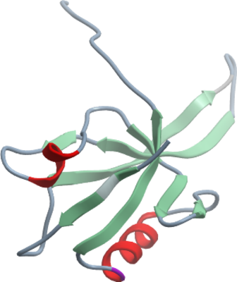
Crystallisation: Crystals grew from a 2:1 ratio of protein to precipitant solution (25 w/v PEG_MME_2000, 85 mM Imidazole pH 6.1, 15 mM Imidazole pH 8.1), using the vapour diffusion method. |
Data Collection: Crystals were cryo-protected by equilibration into precipitant solution containing 25% Ethylene glycol, and then flash frozen in liquid nitrogen. Data was collected at the Swiss Light source, beamline X10. |