Entry Clone Source: Origene |
Entry Clone Accession: GI:65508448 |
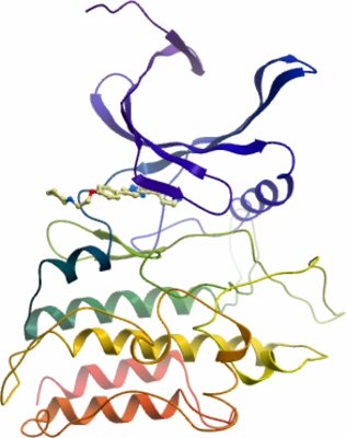
SGC Construct ID: ACVR2A-c046 |
GenBank GI number: gi|4501897 |
Vector: pFB-LIC-Bse. Details [ PDF ]; Sequence [ FASTA ] or [ GenBank ] |
Amplified construct sequence:
TACTTCCAATCCATGCCACTGCAGTT
ATTAGAAGTGAAAGCAAGGGGAAGAT
TTGGTTGTGTCTGGAAAGCCCAGTTG
CTTAACGAATATGTGGCTGTCAAAAT
ATTTCCAATACAGGACAAACAGTCAT
GGCAAAATGAATACGAAGTCTACAGT
TTGCCTGGAATGAAGCATGAGAACAT
ATTACAGTTCATTGGTGCAGAAAAAC
GAGGCACCAGTGTTGATGTGGATCTT
TGGCTGATCACAGCATTTCATGAAAA
GGGTTCACTATCAGACTTTCTTAAGG
CTAATGTGGTCTCTTGGAATGAACTG
TGTCATATTGCAGAAACCATGGCTAG
AGGATTGGCATATTTACATGAGGATA
TACCTGGCCTAAAAGATGGCCACAAA
CCTGCCATATCTCACAGGGACATCAA
AAGTAAAAATGTGCTGTTGAAAAACA
ACCTGACAGCTTGCATTGCTGACTTT
GGGTTGGCCTTAAAATTTGAGGCTGG
CAAGTCTGCAGGCGATACCCATGGAC
AGGTTGGTACCCGGAGGTACATGGCT
CCAGAGGTATTAGAGGGTGCTATAAA
CTTCCAAAGGGATGCATTTTTGAGGA
TAGATATGTATGCCATGGGATTAGTC
CTATGGGAACTGGCTTCTCGCTGTAC
TGCTGCAGATGGACCTGTAGATGAAT
ACATGTTGCCATTTGAGGAGGAAATT
GGCCAGCATCCATCTCTTGAAGACAT
GCAGGAAGTTGTTGTGCATAAAAAAA
AGAGGCCTGTTTTAAGAGATTATTGG
CAGAAACATGCTGGAATGGCAATGCT
CTGTGAAACCATTGAAGAATGTTGGG
ATCACGACGCAGAAGCCAGGTTATCA
GCTGGATGTGTAGGTGAAAGAATTAC
CCAGATGCAGAGACTAACAAATATTT
GACAGTAAAGGTGGATA
|
Final protein sequence (Tag sequence in lowercase):
mghhhhhhssgvdlgtenlyfq^smP
LQLLEVKARGRFGCVWKAQLLNEYVA
VKIFPIQDKQSWQNEYEVYSLPGMKH
ENILQFIGAEKRGTSVDVDLWLITAF
HEKGSLSDFLKANVVSWNELCHIAET
MARGLAYLHEDIPGLKDGHKPAISHR
DIKSKNVLLKNNLTACIADFGLALKF
EAGKSAGDTHGQVGTRRYMAPEVLEG
AINFQRDAFLRIDMYAMGLVLWELAS
RCTAADGPVDEYMLPFEEEIGQHPSL
EDMQEVVVHKKKRPVLRDYWQKHAGM
AMLCETIEECWDHDAEARLSAGCVGE
RITQMQRLTNI
^ TEV cleavage site |
Tags and additions: Cleavable N-terminal His6 tag. |
Host: SF9 Spodoptera frugiperda Insect cells.
|
Growth medium, induction protocol: 2L of SF9 cells at a density of 2x10⁶/ml were infected with 10ml of Virus/L. Cells were incubated at 27°C in the shaker incubator and harvested after 48 hours. Cells were harvested by centrifugation at 4500rpm at 4°C for 15min. Cell pellets from each 1L flask were resuspended in 20ml binding buffer, transferred to 50ml tubes, and stored at -20°C.
Binding buffer: 50 mM HEPES, pH 7.5; 500 mM NaCl; 5% Glycerol; 5 mM Imidazole.
Extraction buffer, extraction method: The frozen cells were thawed and protease inhibitor SET V (Calbiochem) were added to the cell suspension at 1:100 dilutions. The cells were lysed by passage 3-4x through an Emulsiflex C3 homogeniser. The cell lysate was spun down by centrifugation at 25,000rpm at 4°C for 1 hour. The supernatant was recovered for purification. |
Column 1: Anion-exchange for Nucleic acid removal with DEAE cellulose (DE52, Whatmann) 10g of resin was suspended in 100ml 0.2 M NaCl, and then applied onto a 2.5 x 20 cm column. The resin was then equilibrated with 50ml binding buffer prior to loading the sample. |
Column 1 Buffer:
Binding buffer: 50 mM HEPES, pH 7.5; 500 mM NaCl; 5% Glycerol; 5 mM Imidazole; 0.5 mM TCEP.
Wash buffer: 50 mM HEPES, pH 7.5; 500 mM NaCl; 5% Glycerol; 25 mM Imidazole; 0.5 mM TCEP. |
Column 1 Procedure: The supernatant was first applied onto the column by gravity flow, which was followed by a wash with 50ml wash buffer amd a subsequent wash with 50ml wash buffer. The column flow-through and wash was directly applied onto a Ni-sepharose column. |
Column 2: Ni-Affinity Chromatography. 4ml of 50% Ni-sepharose slurry was applied onto a 1.5 x 10 cm column. The column was equilibrated with binding buffer (25ml). |
Column 2 Buffers:
Binding buffer: 50 mM HEPES, pH 7.5; 500 mM NaCl; 5% glycerol; 5 mM imidazole; 0.5 mM TCEP.
Wash buffer: 50 mM HEPES, pH 7.5; 500 mM NaCl; 5% glycerol; 25 mM Imidazole; 0.1 mM TCEP.
Elution buffer: 50 mM HEPES, pH 7.5; 500 mM NaCl; 5% glycerol, 50 to 250 mM Imidazole; 0.5 mM TCEP (step elution).
|
Column 2 Procedure: The flow-through and wash fractions from column 1 (DE52) was applied by gravity flow onto the Ni-sepharose column. The bound protein was then washed with 50ml binding buffer and subsequently with 50ml wash buffer. ACVR2A protein was then eluted by applying a step gradient of imidazole - using 5ml portions of elution buffer with increasing concentration of imidazole (50 mM, 100 mM, 150 mM and 250 mM). Elution fractions were analysed by SDS PAGE and the initial 50 and 100 mM imidazole fractions were kept and pooled. 10 mM DTT was added for overnight storage at 4°C. |
Enzymatic treatment: The tag was not cleaved. |
Column 3: Size Exclusion Chromatography. Superdex S200 16/60 HiLoad. |
Column 3 Buffer: 300 mM NaCl; 50 mM HEPES, pH 7.5; 0.5 mM TCEP, pH 7.5. |
Column 3 Procedure: Prior to applying the protein, the S200 16/60 column was washed and equilibrated with gel filtration buffer. The protein was concentrated to 3ml using and Amicon Ultra-15 filter with a 30kDa cut-off. The concentrated was directly applied onto the equilibrated S200 16/60 column, and run at a flow-rate of 1ml/min. Fractions containing the protein were pooled together. |
Protein concentration: Protein was concentrated to 20mg/ml using an Amicon 30kDa cut-off concentrator. |
Mass spectrometry characterization: The purified protein was homogeneous and had an experimental mass of 36410.3 Da. The theoretical expected mass from the construct primary sequence is 36497.9. The difference corresponds to loss of the N-terminal methionine and acetylation of the resulting N-terminus. Mass was determined by LC-MS, using an Agilent LC/MSD TOF system with reversed-phase HPLC coupled to electrospray ionisation and an orthogonal time-of-flight mass analyser. Proteins were desalted prior to mass spectrometry by rapid elution off a C3 column with a gradient of 5-95% isopropanol in water with 0.1% formic acid. |
Crystallisation: Protein was concentrated to 20mg/ml in gel filtration buffer. Dorsomorphin was added to the final sample at a concentration of 1 mM. Crystals were grown at 20°C in 150nl sitting drops mixing 75nl protein solution with 75nl of a reservoir solution containing 28% PEG3350, 0.2 M lithium sulphate, 10% ethylene glycol, 100 mM Tris-HCl pH 8.8. On mounting crystals were cryoprotected with an additional 20% ethylene glycol. |
Data Collection: 1.96Å resolution.
Phasing: Diamond Light Source, station I03, using monochromatic radiation at wavelength 0.9763Å. |