Entry Clone Source: Site-directed mutagenesis |
Entry Clone Accession: n/a |
SGC Construct ID: ACVR1A-c096 |
GenBank GI number: gi|4501895 |
Vector: pFB-LIC-Bse. Details [ PDF ]; Sequence [ FASTA ] or [ GenBank ] |
Amplified construct sequence:
CCATGGGCCACCATCATCATCATCAT
TCTTCTGGTGTAGATCTGGGTACCGA
GAACCTGTACTTCCAATCCATGCAAA
GAACAGTGGCTCGCGATATTACACTG
TTGGAGTGTGTCGGGAAAGGCAGGTA
TGGTGAGGTGTGGAGGGGCAGCTGGC
AAGGGGAAAATGTTGCCGTGAAGATC
TTCTCCTCCCGTGATGAGAAGTCATG
GTTCAGGGAAACGGAATTGTACAACA
CTGTGATGCTGAGGCATGAAAATATC
TTAGGTTTCATTGCTTCAGACATGAC
ATCAAGACACTCCAGTACCCAGCTGT
GGTTAATTACACATTATCATGAAATG
GGATCGTTGTACGACTATCTTCAGCT
TACTACTCTGGATACAGTTAGCTGCC
TTCGAATAGTGCTGTCCATAGCTAGT
GGTCTTGCACATTTGCACATAGAGAT
ATTTGGGACCCAAGGGAAACCAGCCA
TTGCCCATCGAGATTTAAAGAGCAAA
AATATTCTGGTTAAGAAGAATGGACA
GTGTTGCATAGCAGATTTGGGCCTGG
CAGTCATGCATTCCCAGAGCACCAAT
CAGCTTGATGTGGGGAACAATCCCCG
TGTGGGCACCAAGCGCTACATGGCCC
CCGAAGTTCTAGATGAAACCATCCAG
GTGGATTGTTTCGATTCTTATAAAAG
GGTCGATATTTGGGCCTTTGGACTTG
TTTTGTGGGAAGTGGCCAGGCGGATG
GTGAGCAATGGTATAGTGGAGGATTA
CAAGCCACCGTTCTACGATGTGGTTC
CCAATGACCCAAGTTTTGAAGATATG
AGGAAGGTAGTCTGTGTGGATCAACA
AAGGCCAAACATACCCAACAGATGGT
TCTCAGACCCGACATTAACCTCTCTG
GCCAAGCTAATGAAAGAATGCTGGTA
TCAAAATCCATCCGCAAGACTCACAG
CACTGCGTATCAAAAAGACTTTGACC
AAAATTGATTGACAGTAAAGGTGGAT
ACGGATCCGAATTCGAGCTCCGTCGA
CAAGCTT
|
Final protein sequence (Tag sequence in lowercase):
mghhhhhhssgvdlgtenlyfq^smQ
RTVARDITLLECVGKGRYGEVWRGSW
QGENVAVKIFSSRDEKSWFRETELYN
TVMLRHENILGFIASDMTSRHSSTQL
WLITHYHEMGSLYDYLQLTTLDTVSC
LRIVLSIASGLAHLHIEIFGTQGKPA
IAHRDLKSKNILVKKNGQCCIADLGL
AVMHSQSTNQLDVGNNPRVGTKRYMA
PEVLDETIQVDCFDSYKRVDIWAFGL
VLWEVARRMVSNGIVEDYKPPFYDVV
PNDPSFEDMRKVVCVDQQRPNIPNRW
FSDPTLTSLAKLMKECWYQNPSARLT
ALRIKKTLTKID
^ TEV cleavage site
Engineered Q207D mutation in bold and underlined. |
Tags and additions: Cleavable N-terminal His6 tag. |
Host: SF9 Spodoptera frugiperda Insect cells.
|
Growth medium, induction protocol: SF9 cells at a density of 2x10⁶/ml were infected with recombinant ACVR1 baculovirus (virus stock P3; 1ml of virus stock/100ml of cell culture). Cell were shaken at 110rpm at 27°C in an Innova shaker. After 48 hours post-infection the cultures were harvested by centrifugation for 20min at 6000rpm. Cell pellets from each 1L flask were resuspended in 15ml binding buffer. Calbiochem protease inhibitor SET V was added to the cell suspension at a 1:2000 dilution and transferred to 50ml tubes, and stored at -20°C.
Binding buffer: 50 mM HEPES, pH 7.5; 500 mM NaCl; 5% Glycerol; 5 mM Imidazole.
Extraction buffer, extraction method: The frozen cells were thawed and the volume increased to 200ml with binding buffer. The cells were lysed using an Emulsiflex C5 homogeniser. The cell lysate was spun down by centrifugation at 21,000rpm and 4°C for 1 hour. The supernatant was recovered for purification. |
Column 1: Anion-exchange for Nucleic acid removal with DEAE cellulose (DE52, Whatmann) 10g of resin was suspended in 50ml 1 M NaCl, and then applied onto a 2.5 x 20 cm column. The resin was then equilibrated with 50ml binding buffer prior to loading the sample. |
Column 1 Buffer:
Binding buffer: 50 mM HEPES, pH 7.5; 500 mM NaCl; 5% Glycerol; 5 mM Imidazole; 0.1 mM TCEP.
Wash buffer: 50 mM HEPES, pH 7.5; 500 mM NaCl; 5% Glycerol; 25 mM Imidazole; 0.1 mM TCEP. |
Column 1 Procedure: The supernatant was first applied onto the column by gravity flow, which was followed by a wash with 50ml wash buffer. The column flow-through and wash was directly applied onto a Ni-sepharose column. |
Column 2: Ni-Affinity Chromatography. 6ml of 50% Ni-sepharose slurry was applied onto a 1.5 x 10 cm column. The column was equilibrated with binding buffer (25ml). |
Column 2 Buffers:
Binding buffer: 50 mM HEPES, pH 7.5; 500 mM NaCl; 5% glycerol; 5 mM imidazole; 0.1 mM TCEP.
Wash buffer: 50 mM HEPES, pH 7.5; 500 mM NaCl; 5% glycerol; 25 mM Imidazole; 0.1 mM TCEP.
Elution buffer: 50 mM HEPES, pH 7.5; 500 mM NaCl; 5% glycerol, 50 to 250 mM Imidazole; 0.1 mM TCEP (step elution).
|
Column 2 Procedure: The flow-through from column 1 (DE52) was applied by gravity flow onto the Ni-sepharose column. The bound protein was eluted by applying a step gradient of imidazole - using 10ml portions of elution buffer with increasing concentration of imidazole (50 mM, 100 mM, 150 mM and 250 mM). |
Enzymatic treatment: 0.1mg of TEV protease was added to the Ni-eluted protein to remove the tag. |
Column 3: Size Exclusion Chromatography. Superdex S200 16/60 HiLoad. |
Column 3 Buffer: 300 mM NaCl; 50 mM HEPES, pH 7.5; 0.5 mM TCEP. |
Column 3 Procedure: Prior to applying the protein, the S200 16/60 column was washed and equilibrated with gel filtration buffer. The protein was concentrated to 5ml using and Amicon Ultra-15 filter with a 10kDa cut-off. The concentrated was directly applied onto the equilibrated S200 16/60 column, and run at a flow-rate of 1ml/min. The protein was eluted at 90-108ml. Fractions containing the protein were pooled together. |
Column 4: Ni-Affinity Chromatography. 0.75ml of 50% Ni-sepharose slurry was applied onto 1.5 x 10 cm column. The column was equilibrated with binding buffer (15ml). |
Column 4 Buffer:
Binding buffer: 50 mM HEPES, pH 7.5; 500 mM NaCl; 5% Glycerol; 5 mM Imidazole; 0.1 mM TCEP.
Wash buffer: 50 mM HEPES, pH 7.5; 500 mM NaCl; 5% Glycerol; 25 mM; 0.1 mM TCEP.
Elution buffer: 50 mM HEPES, pH 7.5; 500 mM NaCl; 5% Glycerol; 250 mM Imidazole; 0.1 mM TCEP. |
Column 4 Procedure: The cleaved protein was passed through the column followed by 3ml binding buffer. It was then washed with 8ml wash buffer. Anything remaining bound to the column was eluted with 15ml elution buffer. |
Mass spectrometry characterization: The purified protein was homogeneous and had an experimental mass of 34494.5 (after TEV cleavage), closely matching the expected mass 34492.7. Mass was determined by LC-MS, using an Agilent LC/MSD TOF system with reversed-phase HPLC coupled to electrospray ionisation and an orthogonal time-of-flight mass analyser. Proteins were desalted prior to mass spectrometry by rapid elution off a C3 column with a gradient of 5-95% isopropanol in water with 0.1% formic acid. |
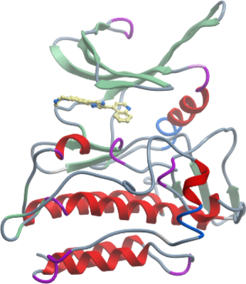
Crystallisation: Protein was buffered in 50 mM HEPES pH 7.5, 300 mM NaCl, 10 mM DTT and 10 mM L-arginine, 10 mM L-glutamate. The protein was concentrated to 10mg/ml (calculated using an extinction co-efficient of 58900) in the presence of the inhibitor LDN-193189 (1mM final concentration). Crystals were grown at 20°C in 150nl sitting drops mixing 75nl protein solution with 75nl of a reservoir solution containing 20% PEG 3350 and 0.20M (NH4)2H(cit) pH 5.0. On mounting crystals were cryoprotected with mother liquor plus 20% ethylene glycol before transfer to liquid nitrogen. |
Data Collection: 1.82Å resolution.
Phasing: Diamond Light Source, station I03. |