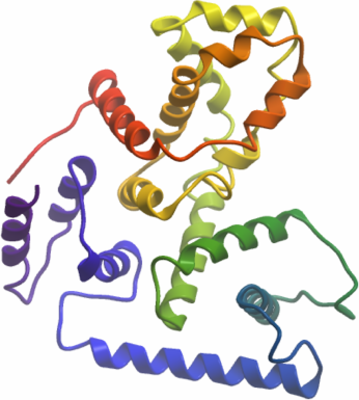
TBC1D7
PDB:3QWL
Revision
Revision Type:created
Revised by:created
Revision Date:created
Entry Clone Accession:BC007054
Entry Clone Source:MGC AU41-B9
SGC Clone Accession:HPC055-E01
Tag:N-terminal His6-tag, removed before crystallization
Host:BL21-V2R-pRARE2
Construct
Prelude:TBC1D7:M1-S293
Sequence:gMTEDSQRNFRSVYYEKVGFRGVEEKKSLEILLKDDRLDTEKLCTFSQRFPLPSMYRALVWKVLLGILPPHHESHAKVMMYRKEQYLDVLHALKVVRFVSDATPQAEVYLRMYQLESGKLPRSPSFPLEPDDEVFLAIAKAMEEMVEDSVDCYWITRRFVNQLNTKYRDSLPQLPKAFEQYLNLEDGRLLTHLRMCSAAPKLPYDLWFKRCFAGCLPESSLQRVWDKVVSGSCKILVFVAVEILLTFKIKVMALNSAEKITKFLENIPQDSSDAIVSKAIDLWHKHCGTPVHSS
Vector:pET28-MHL
Growth
Medium:Antibiotics:Procedure:LEX Bubbling. The target protein was expressed in E. coli by inoculating each 50 mL of overnight culture grown in Luria-Bertani medium into a 2 L of Terrific Broth medium in the presence of 50 mg/mL kanamycin and 30 mg/mL chloramphenicol at 37 degree. When OD600 reached ~3.0, the temperature of the medium was lowered to 15 degree and the culture was induced with 1 mM IPTG. The cells were allowed to grow overnight before harvested and flash frozen in liquid nitrogen and stored at -80 degree.
Selenomethionine labeling of the protein used the M9 SeMEt growth media kit from Medicilon following the manufacturer's instructions.
Purification
ProcedureThe lysate was centrifuged at 15,000 rpm for 45 minutes and the supernatant was mixed with 4 mL 50% flurry of Ni-NTA beads and incubated at 4 degree on a rotary shaker for one and a half hour. The mixture was then centrifuged at 2300 rpm for 5 min and the supernatant discarded. The beads were then washed with 50 mL binding buffer containing 25 mM and 50 mM Imidazole. Bound proteins were eluted using 10 mL elution buffer. The His6 tag was removed by digesting with TEV protease at 1:10 (w/w TEV:protein) ratio overnight in dialysis against gel filtration buffer. The protein was further purified using a Superdex-75 gel filtration column pre-equilibrated with gel filtration buffer. Fractions containing the target protein were pooled and concentrated using Amicon Ultra-15 centrifugal filter (mwco 10 kDa). The purity of the preparation is tested by SDS-PAGE to be greater than 90%.
Extraction
ProcedureFrozen cells from 4L TB culture were thawed and resuspended in 5mL extraction buffer per gram of cell pellets with freshly added 0.2% NP-40, and supplemented with protease inhibitor 1 mM PMSF, and 10U/mL benzonase (Sigma Catalog # E1014, 250U/microL). The cells were lysed using sonication for 8 min at 110 W, 10 sec on/10 sec off duty cycle.
Concentration:15 mg/mL
LigandMassSpec:uncut version native protein expected 36164.8, measured 36166.3
SeMet cut version, expected 34450.66 (for 9 SeMet for +47.94 each) , measured 34455.2
Crystallization:In situ proteolysis were used to get crystals for structure determination. One volume of Dispase I from Bacillus polymyxa (Sigma-Aldrich, Cat. D4818) dissolved in a solution of 10 mM Tris-HCl at pH 7.5 with 100 mM NaCl at a final concentration of 1 mg/mL was mixed with 10 volume protein stock solution (15 mg/mL) right before setting up crystallization. Crystal was initially obtained from SGC-I screen condition A07, D07, and F07.Crystal used for structure refinement was grown in SGC-I screen condition F07, i.e. in 20% PEG 1500, 0.2M MgCl2, 0.1M Tris pH 8.5, in sitting drop setup, using 0.5 uL protein (protein/dispase mix) + 0.5 uL well solution against 100 uL reservoir buffer at room temperature.
Crystal used for phasing was SeMet labelled, grown in SGC-I screen condition A07, i.e. in 30% PEG 1500, 0.2M NaCl, 0.1M HEPES pH 7.5, in sitting drop setup, using 0.5uL protein (protein/dispase mix) + 0.5uL well solution against 100 uL reservoir buffer at room temperature.
Crystals grow to a mountable size within two days.Cryoprotectant used paratone-N
NMR Spectroscopy:
Data Collection:
Data Processing: