Entry Clone Source: Synthetic |
Entry Clone Accession: GI:11067749 |
Vector: pNIC28-Bsa4 Details [PDF ]; Sequence [ FASTA ] or [ GenBank ] |
Amplified DNA sequence:
CATATGCACCATCATCATCATCATTC
TTCTGGTGTAGATCTGGGTACCGAGA
ACCTGTACTTCCAATCCATGCCGGAG
GTCTCCAACCCCAGCAAGCCCGGCCG
CAAGACCAACCAGCTGCAGTACATGC
AGAATGTGGTGGTGAAGACGCTCTGG
AAACACCAGTTCGCCTGGCCCTTCTA
CCAGCCCGTGGACGCAATCAAATTGA
ACCTGCCGGATTATCATAAAATAATT
AAAAACCCAATGGATATGGGGACTAT
TAAGAAGAGACTAGAAAATAATTATT
ATTGGAGTGCAAGCGAATGTATGCAG
GACTTCAACACCATGTTTACAAATTG
TTACATTTATAACAAGCCCACAGATG
ACATAGTGCTAATGGCCCAAGCTTTA
GAGAAAATTTTTCTACAAAAAGTGGC
CCAGATGCCCCAAGAGGAATGACAGT
AAAGGTGGATACGGATCCGAA
|
Final protein sequence (Tag sequence in lowercase):
mhhhhhhssgvdlgtenlyfq^smPE
VSNPSKPGRKTNQLQYMQNVVVKTLW
KHQFAWPFYQPVDAIKLNLPDYHKII
KNPMDMGTIKKRLENNYYWSASECMQ
DFNTMFTNCYIYNKPTDDIVLMAQAL
EKIFLQKVAQMPQEE
^ TEV cleavage site |
Tags and additions: Cleavable N-terminal His6 tag. |
Host: BL21 (DE3)R3-pRARE2 (Phage resistant strain)
|
Growth medium, induction protocol: 10 ml from a 50 ml overnight culture containing 50 µg/ml kanamycin and 34 µg/ml chloramphenicol were used to inoculate each of two 1 liter cultures of TB containing 50 µg/ml kanamycin and 34 µg/ml chloramphenicol. Cultures were grown at 37°C until the OD600 reached ~2.5 then the temperature was adjusted to 18°C. Expression was induced overnight using 0.1 mM IPTG at an OD600 of 3.0. The cells were collected by centrifugation and the pellet re-suspended in binding buffer and frozen.
Binding buffer: 50 mM HEPES pH 7.5; 500 mM NaCl; 10 mM imidazole, 5% glycerol.
Extraction buffer, extraction method: Frozen pellets were thawed and fresh 0.5 mM TCEP, 1 mM PMSF added to the lysate. Cells were lysed using sonication. DNA was precipitated with 0.15% PEI. The lysate was centrifuged at 17,000 rpm for 60 minutes and the supernatant collected for purification. |
Column 1: Ni-affinity. Ni-sepharose (Amersham), 5 ml of 50% slurry in 1.5 x 10 cm column, washed with binding buffer |
Column 1 Buffers:
Binding buffer: 50 mM HEPES pH 7.5, 500 mM NaCl, 5 mM imidazole, 5% glycerol
Wash buffer: 50 mM HEPES pH 7.5, 500 mM NaCl, 30 mM Imidazole, 5% glycerol
Elution buffer: 50 mM HEPES pH 7.5, 500 mM NaCl, 5% glycerol, 50 to 250 mM Imidazole (step elution). |
Column 1 Procedure: The supernatant was loaded by gravity flow on the Ni-sepharose column. The column was then washed first with 30 ml of binding buffer then with 30 ml wash buffer at gravity flow. The protein was eluted by gravity flow by applying 5ml portions of elution buffer with increasing concentration of imidazole (50 mM, 100 mM, 150 mM, and 2x 250 mM); fractions were collected until essentially all protein was eluted. |
Enzymatic treatment: The N-terminal His tag was cleaved by treatment with TEV protease, overnight. |
Column 2: Size Exclusion Chromatography; Superdex S75 16/60 HiLoad |
Column 2 Buffers:
Buffer: 10 mM HEPES, pH 7.5; 500 mM NaCl, 5% glycerol. |
Column 2 Procedure: The protein was concentrated and applied to an S75 16/60 HiLoad gel filtration column equilibrated in 10 mM HEPES, pH 7.5; 500mM NaCl, 5% glycerol using an ÄKTAexpress system. |
Protein concentration: The protein was concentrated to 8.7 mg/ml using an Amicon 3 kDa cut-off concentrator. |
Mass spectrometry characterization:
Measured mass:: 14570 Da
LC- ESI -MS TOF gave a measured mass of 14570 Da for this construct as predicted from the sequence of this protein |
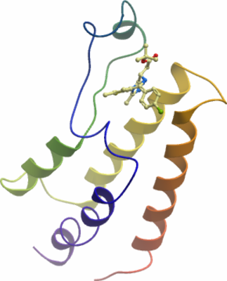
Crystallisation: Protein buffer was exchanged to 10 mM HEPES pH7.5 and 300 mM NaCl. Crystals were grown at 4°C in 300 nl sitting drops from a 1:1 ratio of protein to reservoir solution containing 14% isopropanol, 0.14M CaCl2, 30% glycerol, 0.7M acetate pH 4.6. |
Data collection:
Resolution: 2.10Å
Crystals were cryo-protected using the well solution and flash frozen in liquid nitrogen.
Diffraction data were collected from a single crystal on an Rigaku FRE at a single wavelength of 1.52 Å and the structure was refined to 2.10 Å.
Phasing: The structure was solved by molecular replacement using an ensemble of known bromodomain structures as a starting model |