CDKL3
PDB:3ZDU
Revision
Revision Type:created
Revised by:created
Revision Date:created
Entry Clone Accession:GI: 166064026
Entry Clone Source:Site-directed mutagenesis
SGC Clone Accession:CDKL3A-c014
Tag:MGHHHHHHSSGVDLGTENLYFQ*S, TEV-cleavable (*) C-terminal hexahistidine tag.
Host:SF9 Spodoptera frugiperda Insect cells
Construct
Prelude:
Sequence:MGHHHHHHSSGVDLGTENLYFQSMEMYETLGKVGEGSYGTVMKCKHKNTGQIVAIKIFYERPEQSVNKIAMREIKFLKQFHHENLVNLIEVFRQKKKIHLVFEFIDHTVLDELQHYCHGLESKRLRKYLFQILRAIDYLHSNNIIHRDIKPENILVSQSGITKLCDFGFARTLAAPGDIYDDEVATRWYRAPELVLKDTSYGKPVDIWALGCMIIEMATGNPYLPSSSDLDLLHKIVLKVGNLSPHLQNIFSKSPIFAGVVLPQVQHPKNARKKYPKLNGLLADIVHACLQIDPADRISSSDLLHHEYFTRDGFIEKFMPELKAKLLQEAKVNSLIKPKESSKENEL
Vector:pFB-LIC-Bse
Growth
Medium:2 L of SF9 cells at a density of 2million/ml were infected with 10ml of Virus/L.Cells were incubated at 27°C in the shaker incubator and harvested after 48 hours. Cells were harvested by centrifugation at 900xG at 4°C for 15 min. Cell pellets from each flask (1l volume) were resuspended in 15ml binding buffer, transferred to 50ml tubes, and stored at -20°C.
Antibiotics:
Procedure:
Purification
Buffers
Procedure
Extraction
Buffers
Binding buffer: 50 mM HEPES, pH 7.5; 500 mM NaCl; 5 mM Imidazole; 5% Glycerol.
Procedure
Extraction buffer, extraction method: The frozen cells were thawed and protease inhibitor SET V (Calbiochem) added to the cell suspension at 1:1000 dilution. The cells were lysed by ultrasonication on ice over 15 min with the sonicator pulsing ON for 5 sec and OFF for 10 sec. The cell lysate was clarified by centrifugation at 21,000 rpm at 4°C for 1 h and filtered using syringe filters with a 1.2μm pore size.Column 1: Ni-Affinity Chromatography  2ml Ni-sepharose slurry applied to a 1.5 x 10 cm column.Buffers: Binding buffer: 50mM HEPES, pH 7.5; 500mM NaCl; 5% Glycerol; 5mM Imidazole Wash buffer: 50mM HEPES, pH 7.5; 500mM NaCl; 5% Glycerol; 30mM Imidazole Elution buffer: 50mM HEPES, pH 7.5; 500mM NaCl; 5% Glycerol; 50 to 500mM ImidazoleProcedure: 2ml of 50 % Ni-sepharose slurry (Amersham) was equilibrated in binding buffer and added to the filtered lysate, which was incubated with the Ni-sepharose for 1 hour at 4°C with slow rotation to maximize binding. The lysate was then applied to a 1.5 x 10 cm column by gravity flow. The remaining resin was then washed with 2x50ml binding buffer and 1x30ml wash buffer to remove nonspecifically binding proteins. The bound target protein was eluted by applying a step gradient of imidazole (5 ml fractions of elution buffer supplemented with 50mM and 500mM imidazole). The protein content of collected fractions was visualized using SDS-PAGE and fractions containing CDKL3A were pooled. Enzymatic treatment: TEV protease cleavage. Pooled fractions were treated with TEV protease overnight at 4°C.Column 2: Size Exclusion Chromatography  S75 HiLoad 26/60 Superdex run on ÄKTAprime.Buffer: Gel Filtration buffer: 10mM HEPES pH7.5, 250mM NaCl, 5% Glycerol, 1mM TCEPProcedure: Prior to applying the protein, the S75 26/60 column was washed and equilibrated with gel filtration buffer. Eluted protein from Ni-sepharose column was concentrated to 2ml using an Amicon Ultra-15 filter with a 10kDa cut-off. The concentrated protein was directly applied onto the equilibrated S75 26/60 column, and run at a flow-rate of 2.5 ml/min. 3ml fractions were collected and visualized using SDS-PAGE. Those containing CDKL3A were pooled.
Concentration:The protein was concentrated to a final concentration of 9mg/ml (measured by OD280 based on extinction coefficient 28880) in an Amicon Ultra-15 filter with a 30 kDa cut-off.
Ligand
MassSpec:The purified protein was homogeneous and had an experimental mass of 37291.4 Da. The theoretical expected mass from the construct sequence is 37292.4 Da. Masses were determined by LC-MS, using an Agilent LC/MSD TOF system with reversed-phase HPLC coupled to electrospray ionisation and an orthogonal time-of-flight mass analyser. Proteins were desalted prior to mass spectrometry by rapid elution off a C3 column with a gradient of 5-95% methanol in water with 0.1% formic acid.
Crystallization:Protein at 9mg/ml was buffered in 10mM HEPES, pH 7.5, 250mM NaCl, 1 mM TCEP. 2-[4-({4-[(5-cyclopentyl-1H-pyrazol-3-yl)amino]pyrimidin-2-yl}amino)phenyl]acetonitrile (pan-kinase inhibitor ASC67) was added to the final sample. Crystals were grown at 4°C in 150 nl sitting drops mixing 75 nl protein solution with 75 nl of a reservoir solution containing 0.03M ZnCl, 30% (w/v) PEG6000, 5% ethylene glycol, 0.1M MES pH5.5. On mounting crystals were cryo-protected with an additional 25% ethylene glycol.
NMR Spectroscopy:
Data Collection:Resolution: 2.2 Ã
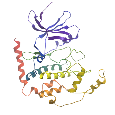
X-ray source: Diamond I04 Crystals of CDKL3A diffracted to a resolution of 2.2 Ã
(scaled resolution). A full dataset was collected at 100 K on Diamond Light Source beamline I04. Crystals belonged to the Trigonal space group P3121 with unit-cell parameters a=63.9 Ã
b=63.9 Ã
c=163.6 Ã
, α=90° β= 90 γ= 120°. Only one molecule was present in the asymmetric unit. Data were indexed and integrated using iMOSFLM and scaled using AIMLESS. Phases were found using molecular replacement in PHASER. PHENIX.SCULPTOR was used to optimize PDB entries 4AAA and 4AGU for use as an ensemble search model. The structure was built using PHENIX.AUTOBUILD and then refined and modified using alternate rounds of REFMAC5 and COOT. The final model was validated using the PHENIX validation tools.
Data Processing: