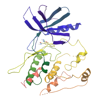
TYK2
PDB:3ZON
Revision
Revision Type:created
Revised by:created
Revision Date:created
Entry Clone Accession:
Entry Clone Source:Mammalian Gene Collection (IMAGE Consortium Clone ID 4591726).
SGC Clone Accession:
Tag:N-terminal, TEV cleavable hexahistidine tag
Host:
Construct
Prelude:
Sequence:MGHHHHHHSSGVDLGTENLYFQSMGDDCFSLRRCCLPQPGETSNLIIMRGARASPRTLNLSQLSFHRVDQKEITQLSHLGQGTRTNVYEGRLRVEGSGDPEEGKMDDEDPLVPGRDRGQELRVVLKVLDPSHHDIALAFYETASLMSQVSHTHLAFVHGVCVRGPENIMVTEYVEHGPLDVWLRRERGHVPMAWKMVVAQQLASALSYLENKNLVHGNVCGRNILLARLGLAEGTSPFIKLSDPGVGLGALSREERVERIPWLAPECLPGGANSLSTAMDKWGFGATLLEICFDGEAPLQSRSPSEKEHFYQRQHRLPEPSCPQLATLTSQCLTYEPTQRPSFRTILRDLTRLQPHN The N-terminal residues, MGHHHHHHSSGVDLGTENLYFQSM, derive from the vector.
Vector:pFB-LIC-Bse
Growth
Medium:
Antibiotics:
Procedure:
Purification
Procedure
Cell Lysis: The resuspended cell pellet was thawed and lysed by sonication. PEI (polyethyleneimine) was added to a final concentration of 0.1 %. The cell debris and precipitated DNA were spun down.
Lysis Buffer: 50 mM Tris pH 7.8, 200 mM NaCl, 20 mM Imidazole, 0.5 mM TCEP, 1:1000 dilution of Sigma protease inhibitor cocktail.
Purification:
Column 1: 6 ml of Ni-Sepharose in a 2 cm diameter gravity flow column.
Column 1 Buffers:
Binding Buffer:50 mM Tris pH 7.8, 200 mM NaCl, 20 mM Imidazole, 0.5 mM TCEP.
Wash Buffer 1:As Binding Buffer except 1 M NaCl and 40 mM imidazole.
Wash Buffer 2:As Binding Buffer except 60 mM imidazole.
Elution Buffer:As Binding Buffer except 250 mM imidazole.
Column 1 Procedure:The clarified supernatant was passed through the column. The column was washed with 50 ml of Binding Buffer (Wash 1) and 50 ml of Wash Buffer 1 (Wash 2) and 40 ml of Wash Buffer 2 (Wash 3). 36 ml of Elute Buffer was passed through to elute the protein. The Wash 3 and Elute fractions were combined and TEV protease was added, The sample was left at 4C overnight.
Column 2: S200 16/60 Gel Filtration (GE Healthcare)
Column 2 Buffers:
GF Buffer:20 mM Tris pH 7.8, 200 mM NaCl, 0.5 mM TCEP
Column 2 Procedure:The eluted protein was concentrated to 5 ml volume and injected onto the column.
Column 3: 1 ml of Ni-Sepharose in a 0.5 cm diameter gravity flow column
Column 3 Procedure:The pooled fractions from gel filtration were passed through the column (pre-equilibrated in GF Buffer) followed by 5 ml of addition GF Buffer. The resin was eluted with 5 ml of GF Buffer containing 10 mM, 20 mM, 30 mM and finally 40 mM imidazole.
Column 4: S200 16/60 Gel Filtration (GE Healthcare)
Column 4 Buffers:
GF Buffer:20 mM Tris pH 7.8, 200 mM NaCl, 0.5 mM TCEP
Column 4 Procedure:The flow-through, 10 mM, 20 mM, 30 mM and 40
mM elutions from column 3 were combined, concentrated, and injected onto the column.
Extraction
Procedure
The TYK2A protein was expressed using baculovirus infected Sf9 cells. Cells were infected at a density of 2,200,000 cells/ml for 48h.
Cell harvest:Cells were spun at 1000x g for 15 mins and the pellets resuspended in Lysis Buffer and then frozen at -20°C.
Concentration:The fractions containing TYK2A were combined. Compound K00238 (IKK-2 Inhibitor VI) was added and the sample concentrated to 8.5 mg/ml (measured by 280 nm absorbance).
Ligand
MassSpec:Expected: 37357.6Observed: 37371.4
Crystallization:Crystals grew in 300 nL drops from a 2 :1 ratio of protein and precipitant solution (0.1M MES pH 6.5 -- 12%(w/v) PEG 20000), using the vapour diffusion method.
NMR Spectroscopy:
Data Collection:Crystals were cryo-protected by equilibration into precipitant solution containing 25% ethylene glycol, and then flash frozen in liquid nitrogen. Data was collected at Diamond, beamline I03.
Data Processing: