Entry Clone Source: Ordered-synthetic |
Entry Clone Accession: Codon-optimized |
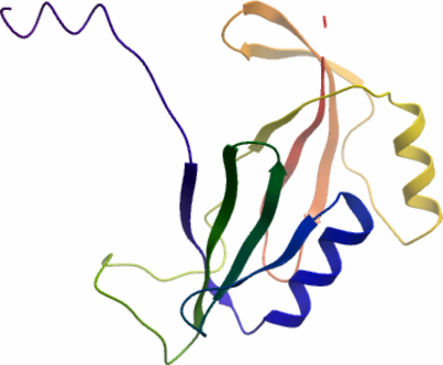
SGC Construct ID: GLOD5A-c102 |
GenBank GI number: gi|122937414 |
Vector: pNIC28-Bsa4. Details [PDF ]; Sequence [ FASTA ] or [ GenBank ] |
Amplified construct sequence:
CATATGCACCATCATCATCATCATTC
TTCTGGTGTAGATCTGGGTACCGAGA
ACCTGTACTTCCAATCCATGCTGATT
CGTCGCCTGGATCATATCGTGATGAC
CGTTAAAAGCATCAAAGACACCACGA
TGTTCTACTCTAAAATCCTGGGCATG
GAAGTTATGACGTTCAAAGAAGATCG
TAAAGCCCTGTGTTTTGGCGACCAGA
AATTCAACCTGCATGAAGTTGGTAAA
GAATTTGAACCGAAAGCAGCACACCC
GGTCCCGGGCAGTCTGGATATTTGCC
TGATCACCGAAGTGCCGCTGGAAGAA
ATGATTCAACACCTGAAAGCATGTGA
CGTCCCGATCGAAGAAGGTCCGGTGC
CGCGTACGGGTGCTAAAGGTCCGATT
ATGAGCATTTATTTTCGTGACCCGGA
CCGTAACCTGATTGAAGTCTCTAACT
ACTGACAGTAAAGGTGGATACGGATC
CGAA
|
Final protein sequence (Tag sequence in lowercase):
mhhhhhhssgvdlgtenlyfq^smLI
RRLDHIVMTVKSIKDTTMFYSKILGM
EVMTFKEDRKALCFGDQKFNLHEVGK
EFEPKAAHPVPGSLDICLITEVPLEE
MIQHLKACDVPIEEGPVPRTGAKGPI
MSIYFRDPDRNLIEVSNY
^ TEV cleavage site |
Tags and additions: Cleavable N-terminal His6 tag. |
Host: BL21 (DE3)R3-pRARE2 (Phage resistant strain).
|
Growth medium, induction protocol: The construct DNA was transformed into competent cells of the expression strain by a standard heat shock procedure. One colony from the transformation was used to inoculate 1ml of TB media containing 50µg/ml kanamycin and 34 µg/ml chloramphenicol, which was placed in a 37°C shaker overnight. The next day glycerol stocks were prepared from this overnight culture. A glycerol stock was used to inoculate 60ml of TB media containing 50µg/ml kanamycin and 34µg/ml chloramphenicol, which was placed in a 37°C shaker overnight. The next day this starter culture was used to inoculate 6L of TB media (9ml starter culture used per 1L) containing 50µg/ml kanamycin. When the OD600 reached approximately 1.0 the temperature was reduced to 18°C and after a further 30 minutes the cells were induced by the addition of 0.1 mM IPTG. The expression was continued overnight. Cells were harvested by centrifugation at 6000 x g after which the supernatant was poured out and the cell pellet either placed in a -80°C freezer or used directly for purification.
Lysis buffer: 50 mM HEPES, pH 7.4; 500 mM NaCl; 10 mM Imidazole; 5% Glycerol; 0.5 mM TCEP; 1 tablet per 50ml protease inhibitor cocktail EDTA-free (Roche).
Extraction buffer, extraction method: Cell pellets were dissolved in approximately 200ml lysis buffer and broken by passing through the homogeniser (x6) at a constant pressure of 15KPa. The cell debris was pelleted at 16,000rpm and the supernatant used for further purification. |
Column 1: Ni-NTA (5.0ml volume in a gravity-flow column). |
Column 1 Buffers:
Binding buffer: 50 mM HEPES, pH 7.4; 500 mM NaCl; 5% glycerol; 10 mM Imidazole; 0.5 mM TCEP.
Wash buffer: 50 mM HEPES, pH 7.4; 500 mM NaCl; 5% glycerol; 40 mM Imidazole; 0.5 mM TCEP.
Elution buffer: 50 mM HEPES, pH 7.4; 500 mM NaCl; 5% glycerol; 250 mM Imidazole; 0.5 mM TCEP. |
Column 1 Procedure: The clarified cell extract was incubated with 5.0ml of Ni-NTA pre-equilibrated with lysis buffer for 1 hour at 4°C with rotation after which it was passed through a glass column. The column was then washed with 50ml Binding Buffer (2 x 25ml) and 50ml Wash Buffer (2 x 25ml). The protein was eluted with 50ml of Elution Buffer in 5 x 5 ml fractions. |
Column 2: Superdex S75 16/60 Gel Filtration. |
GF Buffer: 10 mM HEPES, pH 7.4; 500 mM NaCl; 5% glycerol; 0.5 mM TCEP.
|
Column 2 Procedure: Elution fraction from Ni-NTA column (5ml) was applied directly to the GF column (pre-equilibrated in GF Buffer) at 1.0ml/min. 1.0ml fractions were collected. |
Enzymatic treatment: The N-terminal His tag was cleaved by treatment with TEV protease, overnight. |
Column 3: 1ml Resource Q Cation Exchange |
Column 3 Buffers:
Buffer A
: 50 mM HEPES, pH 7.5; 50 mM NaCl.
Buffer B
: 50 mM HEPES, pH 7.5; 2 M NaCl. |
Column 3 Procedure: Pooled fractions giving a total approximate volume of 12ml were then diluted to 100 ml using Buffer A and injected into a 1ml Resource S column. Protein was eluted using a linear gradient of 0-100% Buffer B over 35 column volumes at 1ml/min. 1.0ml fractions were collected. |
Protein concentration: Fractions containing protein were pooled and concentrated to 14mg/ml using a 10kDa mwco concentrator. |
Mass spectrometry characterization: After TEV protease digestion:
Measured mass: 16434.5Da
Expected mass: 16817.5Da
|
Crystallisation: Crystals were grown by vapour diffusion in sitting drop at 20°C. A sitting drop consisting of 100nl protein and 50nl well solution was equilibrated against well solution containing 2 M ammonium sulfate, 20% (w/v) PEG400 and 0.1 M HEPES, pH 7.0. Crystals were mounted in the presence of 20% (v/v) PEG400 and flash-cooled in liquid nitrogen. |
Data collection:
Resolution: 1.50Å.
X-ray source: Diamond Light Source beamline I03. |