Entry Clone Source: MGC |
Entry Clone Accession: IMAGE:2960155 |
SGC Construct ID: ELAC1A-c005 |
GenBank GI number: gi|8922122 |
Vector: pNIC-CTHF. Details [ PDF ]; Sequence [ FASTA ] or [ GenBank ] |
Amplified construct sequence:
ATGGATGTGACATTCCTGGGGACGGG
TGCAGCATACCCATCTCCAACCCGGG
GTGCCTCTGCTGTGGTCCTTCGGTGT
GAAGGCGAGTGCTGGCTCTTTGACTG
TGGGGAGGGAACACAGACACAGCTTA
TGAAAAGCCAACTTAAAGCAGGGAGA
ATTACCAAGATCTTCATCACACACCT
TCATGGAGACCATTTCTTTGGCCTTC
CTGGGCTCCTCTGCACAATCAGCCTG
CAGAGTGGCTCCATGGTGTCCAAACA
GCCTATTGAAATCTATGGCCCTGTAG
GGCTTCGGGACTTTATCTGGCGAACC
ATGGAACTCTCTCACACGGAGCTGGT
CTTCCATTATGTGGTTCATGAACTGG
TTCCTACAGCAGATCAATGTCCTGCA
GAAGAACTAAAAGAATTTGCGCATGT
GAATAGAGCAGACAGTCCTCCCAAAG
AGGAACAAGGAAGAACTATCCTGTTA
GACTCAGAAGAAAACTCATACCTTCT
GTTTGATGATGAACAATTTGTTGTAA
AAGCATTTCGCCTCTTTCACAGAATT
CCCTCATTTGGGTTTTCAGTCGTGGA
AAAGAAACGCCCAGGTAAACTCAATG
CACAGAAACTTAAAGACCTTGGTGTT
CCACCAGGTCCTGCCTATGGGAAGCT
GAAAAATGGAATTTCTGTTGTTCTGG
AAAATGGGGTTACAATTTCTCCCCAA
GATGTCTTAAAAAAGCCTATTGTTGG
AAGAAAAATCTGCATATTGGGTGACT
GCTCTGGGGTTGTGGGTGATGGAGGA
GTAAAACTGTGCTTTGAAGCAGACCT
GTTGATCCACGAAGCAACCCTGGATG
ATGCCCAGATGGACAAAGCAAAGGAG
CATGGCCACAGCACACCACAGATGGC
AGCAACATTTGCAAAGTTGTGCCGTG
CAAAGAGGCTGGTTCTGACTCACTTC
AGTCAGAGGTACAAACCAGTTGCCTT
GGCCAGAGAAGGAGAAACAGATGGCA
TTGCAGAACTAAAAAAGCAAGCTGAA
TCAGTGTTAGATCTCCAAGAAGTGAC
TCTAGCAGAAGATTTTATGGTGATAA
GCATTCCAATCAAGAAAGCAGAGAAC
CTCTACTTCCAATCGCACCATCATCA
CCACCATGATTACAAGGATGACGACG
ATAAGTGA
|
Final protein sequence (Tag sequence in lowercase):
MDVTFLGTGAAYPSPTRGASAVVLRC
EGECWLFDCGEGTQTQLMKSQLKAGR
ITKIFITHLHGDHFFGLPGLLCTISL
QSGSMVSKQPIEIYGPVGLRDFIWRT
MELSHTELVFHYVVHELVPTADQCPA
EELKEFAHVNRADSPPKEEQGRTILL
DSEENSYLLFDDEQFVVKAFRLFHRI
PSFGFSVVEKKRPGKLNAQKLKDLGV
PPGPAYGKLKNGISVVLENGVTISPQ
DVLKKPIVGRKICILGDCSGVVGDGG
VKLCFEADLLIHEATLDDAQMDKAKE
HGHSTPQMAATFAKLCRAKRLVLTHF
SQRYKPVALAREGETDGIAELKKQAE
SVLDLQEVTLAEDFMVISIPIKKaen
lyfq^shhhhhhdykddddk
^ TEV cleavage site |
Tags and additions: C-terminal TEV-cleavable His6 tag. |
Host: BL21 (DE3)R3-pRARE2.
|
Growth medium, induction protocol: The expression plasmid was transformed into the host strain and plated on LB-agar containing 50µg/ml kanamycin and 34µg/ml chloramphenicol. Several colonies were combined to inoculate a 1ml culture in TB (+ 50µg/ml kanamycin, 34µg/ml chloramphenicol). The culture was grown overnight, glycerol was added to 15% v/v (from a 60% stock), and the resulting glycerol stock was frozen at -80°C in 100µl aliquots. A loopful of cells from the glycerol stock was inoculated into 20ml of TB medium containing 50µg/ml kanamycin and 34µg/ml chloramphenicol and grown overnight at 37°C. 2x1L TB medium (containing 50µg/ml kanamycin) were each inoculated with 10ml of the overnight culture and grown in 2.5L UltraYield baffled flasks until OD600 of 3.0. Cells were cooled to 18°C, IPTG added to 0.1 mM and growth continued at 18°C overnight. The cells were collected by centrifugation then the pellets were scraped out and transferred to 50ml Falcon tubes and frozen at -80°C.
Lysis buffer: 25 mM Tris, pH 8.0; 500 mM NaCl; 5 mM Imidazole; 10% Glycerol; 0.5 mM TCEP; 1x Protease Inhibitors Cocktail Set VII (Calbiochem, 1/1000 dilution).
2x lysis buffer contains the same components at double concentration.
Extraction buffer, extraction method: Frozen cell pellets (86g) were thawed briefly in a bath of warm water (20 - 37°C) then transferred to ice. One volume (i.e. 1ml for every gram of cells) of 2x lysis buffer was added, followed by 1x lysis buffer to a total volume of 300ml. The cells were resuspended by agitating and disrupted by sonication. Nucleic acids and cell debris were removed by adding 0.15% PEI (polyethyleneimine) from a 5% (w/v, pH 7.5) stock, stirring for 15 minutes, then centrifugation for 1 hour at 25,000g. |
Column 1: Histrap FF 5ml (GE Healthcare) |
Column 1 Buffers:
Affinity buffer
: 50 mM HEPES, pH 7.4; 500 mM NaCl; 5% glycerol; 5 mM Imidazole; 0.5 mM TCEP.
Wash buffer
: 50 mM HEPES, pH 7.4; 500 mM NaCl; 5% glycerol; 30 mM Imidazole; 0.5 mM TCEP.
Elution buffer
: 50 mM HEPES, pH 7.4; 500 mM NaCl; 5% glycerol; 250 mM Imidazole; 0.5 mM TCEP. |
Column 1 Procedure: The cell extract was loaded onto the column at 5ml/minute on an ÄKTA-express system (GE Healthcare). The column was then washed with 10 volumes of loading buffer, 10 volumes of wash buffer, then eluted with elution buffer at 4 ml/min. The eluted peak of A280 was automatically collected. |
Column 2: Gel filtration, HiLoad 16/60 Superdex 75, 120 ml |
GF Buffer: 50 mM HEPES, pH 7.4; 0.25 M NaCl; 0.5 mM TCEP. |
Column 2 Procedure: The eluted fraction was loaded and fractionated on the gel filtration column in GF buffer at 1.2 ml/min. 2-ml fractions were collected at the A280 peaks. The fractions were analyzed by SDS-PAGE and relevant fractions were pooled. |
Enzymatic treatment:The protein was incubated with 1:20 mol:mol TEV protease overnight at 4°C. The protein plus TEV was passed through a column containing 0.5ml Ni-NTA pre-equilibrated with GF Buffer. Column was washed 5ml of GF Buffer. Flow-through and wash were pooled.
|
Crystallisation: Crystals were grown by vapor diffusion at 4°C in 150nl sitting drops. Three crystals were used to generate this final crystal structure. A 1.7Å diffracting crystal (Crystal 1) and a derivatised 2.2Å diffracting crystal (Crystal 2) were used in a in a SIRAS experiment, which was used as a molecular replacement model for a different 1.7Å dataset (Crystal 3).
Crystal 1: A crystal was grown by mixing 50nl of protein solution (40mg/mL) and 100nl of precipitant consisting of 15% PEG 3350, 500 µM dGMP, 0.15 M Sodium Nitrate pH 7.0. Crystals appeared in about a week at 20°. Crystals were cryo-protected by bathing the crystal in mother liquor supplemented with 25% ethylene glycol and flash-freezing in liquid nitrogen. This crystal was used to collect a native dataset which was scaled at 1.7Å.
Crystal 2: Another crystal was grown by mixing 50nl of protein solution (40mg/mL) and 100nl of precipitant consisting of 15% PEG 3350, 500µM dGMP, 0.15 M Sodium Nitrate pH 7.0. Crystals appeared in about a week at 20°. The crystal was soaked for 35 minutes with 5 mM Thiomersal, before cryoprotection in mother liquor supplemented with 25% ethylene glycol and flash-freezing in liquid nitrogen. This crystal was scaled at a resolution of 2.2Å.
Crystal 3:
Another crystal was grown by mixing 50nl of protein solution (40mg/mL) and 100nl of precipitant consisting of 15% PEG 3350, 500 µM GMP, 0.2 M Sodium Nitrate pH 7.0. Crystals appeared in about a week at 20°. Crystals were cryo-protected by bathing the crystal in mother liquor supplemented with 25% ethylene glycol and flash-freezing in liquid nitrogen. This crystal was used to collect a native dataset which was scaled at 1.7Å. |
Data collection: Diffraction data for the crystals were collected at the Diamond synchrotron beamline I03, at a wavelength of 0.9795Å for Crystal 1 and 1.00 for Crystal 2 and Crystal 3.
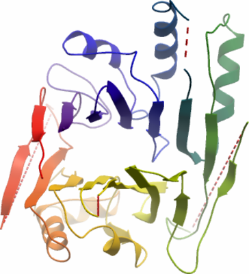
The protein crystallised in space group P21 with two molecules in the asymmetric unit.
6 mercury sites were unambiguously identified using SHELXD, with an anomalous signal extending to ~3.3Å. The derivatised crystal dataset was used with the native dataset in a SIRAS experiment in SHARP, with solvent flattening in SOLOMAN, resulting in an electron density map clearly showing two globular molecules, corresponding to a ELAC1 dimer.
An initial model was built in to this experimentally phased map using the ArpWarp Server. This was refined using Refmac and then used as a search model to phase the dataset from Crystal 3 using Phaser. Iterative rounds of model building in Coot and refinement using Refmac was performed to a final resolution of 1.7Å. |