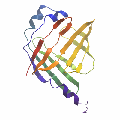
Entry Clone Source: Synthetic |
SGC Construct ID: FABP9A-c002 |
Vector: pNIC-Bsa4 Details [PDF ]; Sequence [ FASTA ] or [ GenBank ] |
Amplified DNA sequence:
CATATGCACCATCATCATCATCATTC
TTCTGGTGTAGATCTGGGTACCGAGA
ACCTGTACTTCCAATCCATGGAGCCC
TTCTTGGGAACCTGGAAGCTGGTCTC
CAGTGAAAACTTTGAGGATTACATGA
AAGAACTGGGAGTGAATTTCGCAGCC
CGGAACATGGCAGGGTTAGTGAAACC
GACAGTAACTATTAGTGTTGATGGGA
AAATGATGACCATAAGAACAGAAAGT
TCTTTCCAGGACACTAAGATCTCCTT
CAAGCTGGGGGAAGAATTTGATGAAA
CTACAGCAGACAACCGGAAAGTAAAG
AGCACCATAACATTAGAGAATGGCTC
AATGATTCACGTCCAAAAATGGCTTG
GCAAAGAGACAACAATCAAAAGAAAA
ATTGTGGATGAAAAAATGGTAGTGGA
ATGTAAAATGAATAATATTGTCAGCA
CCAGAATCTACGAAAAGGTGTGACAG
TAAAGGTGGATACGGATCCGAA
|
Final protein sequence (Tag sequence in lowercase):
mhhhhhhssgvdlgtenlyfq^smEP
FLGTWKLVSSENFEDYMKELGVNFAA
RNMAGLVKPTVTISVDGKMMTIRTES
SFQDTKISFKLGEEFDETTADNRKVK
STITLENGSMIHVQKWLGKETTIKRK
IVDEKMVVECKMNNIVSTRIYEKV
^ TEV cleavage site |
Tags and additions: Tev-cleavable (^) N-terminal hexahistidine tag. |
Host: BL21(DE3)-R3-pRARE2
|
Growth medium, induction protocol: A glycerol stock was used to inoculate 60 ml of TB media containing 50 µg/ml kanamycin and 34 µg/ml chloramphenicol, which was placed in a 37°C shaker overnight. The next day this starter culture was used to inoculate 6L of TB media (9 ml starter culture used per 1L) containing 50 µg/ml kanamycin. When the OD600 reached approximately 1.0 the temperature was reduced to 18°C and after another 30 minutes the cells were induced by the addition of 0.1 mM IPTG. The expression was continued overnight.
Extraction buffer, extraction method: Cell pellets were dissolved in approximately 200ml lysis buffer and broken by passing through the homogeniser at a constant pressure of 15KPa. The cell debris was pelleted at 16,000 RPM and the supernatant used for further purification.
Lysis Buffer: 50 mM Hepes pH 7.4, 500 mM NaCl, 5% Glycerol, 10 mM Imidazole pH 7.4, 0.5 mM TCEP, 1 tablet per 50 ml protease inhibitor cocktail EDTA-free (Roche) |
Column 1: Ni-NTA (5.0 ml in a gravity-flow column). 5ml of 50% Ni-IDA slurry was applied onto a 1.5 x 10cm column. The column was first washed with deionised distilled H2O, and then equilibrated with Binding buffer. |
Column 1 Buffers:
Binding buffer: 50 mM Hepes pH 7.4, 500 mM NaCl, 5% Glycerol, 10 mM Imidazole pH 7.4, 0.5 mM TCEP
Wash buffer: 50 mM Hepes pH 7.4, 500 mM NaCl, 5% Glycerol, 40 mM Imidazole pH 7.4, 0.5 mM TCEP
Elution buffer: 50 mM Hepes pH 7.4, 500 mM NaCl, 5% Glycerol, 250 mM Imidazole pH 7.4, 0.5 mM TCEP |
Column 1 Procedure: The clarified cell extract was incubated with 5.0 ml of Ni-NTA pre-equilibrated with lysis buffer for 1 hour at 4°C with rotation after which it was passed through a glass column. The column was then washed with 50ml Binding Buffer (2 x 25ml) and 50 ml Wash Buffer (2 x25 ml). The protein was eluted with 50 ml of Elution Buffer in 5 x 5 ml fractions. |
Column 2: Superdex S200 16/60 Gel Filtration |
Column 2 Buffers:
Gel Filtration buffer: 10 mM Hepes pH 7.4, 500 mM NaCl, 0.5 mM TCEP, 5% Glycerol |
Column 2 Procedure: Wash buffer fractions 1 and 2 were pooled along with a separate pool of elution fractions 1 and 2, from the Ni-NTA column. Each pool was then concentrate to 5ml and applied directly to the GF column (pre-equilibrated in GF Buffer) at 1.0 ml/min. 1.0 ml fractions were collected. The protein eluted at a volume of between 70 ml and 100 ml for the wash fractions pool and between 40ml and 100 ml for the elution fractions pool. |
Protein concentration: Protein was concentrated to 20 mg/ml using a 10 kDa mwco concentrator. |
Mass spectrometry characterization:
Measured mass: 17574.61 Da
Expected mass: 17546.2 Da |
Crystallisation: Crystals were grown by vapour diffusion in sitting drop at 4°C. A sitting drop consisting of 75 nl protein and 75 nl well solution was equilibrated against well solution containing 1.6M sodium citrate tribasic pH 6.5. Crystals were mounted in the presence of 25% (v/v) ethylene glycol and flash-cooled in liquid nitrogen |
Data collection:
X-ray source: Diamond Light Source Beamline I02
Resolution: 1.7Å |