Growth Medium, Induction Protocol:
The expression plasmid was transformed into the host strain and plated on LB-agar containing 50 µg/ml kanamycin and 34 µg/ml chloramphenicol. Several colonies were combined to inoculate a 1ml culture in TB (+ 50 µg/ml kanamycin, 34 µg/ml chloramphenicol). The culture was grown overnight, glycerol was added to 15% v/v (from a 60% stock), and the resulting glycerol stock was frozen at -80°C in 100 µl aliquots. A loopful of cells from the glycerol stock was inoculated into 20-ml of TB medium containing 50 µg/ml kanamycin and 34 µg/ml chloramphenicol and grown overnight at 37°C. 2x 1L TB medium (containing 50 µg/ml kanamycin) were each inoculated with 10 ml of the overnight culture and grown in 2.5L UltraYield baffled flasks until OD600 of 1.0. Cells were cooled to 18°C, IPTG added to 0.1mM and growth continued at 18°C overnight. The cells were collected by centrifugation then the pellets were scraped out and transferred to 50-ml Falcon tubes and frozen at -80°C.
Cell Extraction: Frozen cell pellets were thawed briefly in a bath of warm water (20 - 37°C), then transferred to ice.. Lysis buffer was added, 2 x w/v. The cells were resuspended by agitating and disrupted by sonication. The lysate was further diluted with 2 volumes of lysis buffer.
Nucleic acids and cell debris were removed by adding 0.15% PEI (polyethyleneimine) from a 5% (w/v) stock, then centrifugation for 1 hour at 25,000 xg.
Lysis buffer: 50 mM HEPES, pH 7.4, 500 mM NaCl, 5 mM imidazole, 5% glycerol,1x Protease Inhibitors Cocktail Set VII (Calbiochem, 1/1000 dilution), 0.5 mM TCEP
PEI (polyethyleneimine) stock: 5% (w/v) pH 7.5, made from 50% solution (sigma P3143), need to adjust pH
|
Column 1: Histrap FF 5 ml (GE Healthcare) |
Column 1 Buffers:
Affinity buffer: 50 mM Hepes, pH 7.5, 500 mM NaCl, 25 mM imidazole, 5% Glycerol, 0.5 mM TCEP
Elution buffer: 50 mM Hepes, pH 7.5, 500 mM NaCl, 250 mM imidazole, 5% Glycerol, 0.5 mM TCEP
|
Column 1 Procedure: The cell extract was loaded onto the column at 5 ml/minute on an AKTA-express system (GE Healthcare). The column was then washed with 10 volumes of loading buffer, 10 volumes of wash buffer, then eluted with elution buffer at 4 ml/min. The eluted peak of A280 was automatically collected.
|
Column 2: Tag cleavage and removal of contaminants |
Column 2 Buffers:
Affinity buffer: 50 mM Hepes, pH 7.5, 500 mM NaCl, 25 mM imidazole, 5% Glycerol, 0.5 mM TCEP
|
Column 2 Procedure: Protein was treated with TEV protease (His6-tagged) overnight at 4C, while being dialysed in wash buffer, using 3000MCO dialysis tubing. The protease and other impurities were removed by passing through 5mL NiNTA column. The column was washed with 10mL of wash buffer which contained a large amount of pure LACTB2A.
The flowthough and wash aliquots analysed by SDS-PAGE and both were pooled.
|
Column 3: Gel filtration, HiLoad 16/60 Superdex Superdex 200, 120 ml. | |
Crystallization: Poorly diffracting crystals (7-11Å) were grown by vapor diffusion at 20°C in 150 nL sitting drops. The drops were prepared by mixing 50 nL of protein solution (33 mg/mL) and 100 nL of precipitant consisting of 0.2 NaBr, 0.1M Bis-Tris-Propane, pH 6.5, 20% PEG 3350 and 10% Ethylene Glycol. These crystals were used to generate seeds for further crystal trials. Several crystals were aliquotted from the crystal drop into 100 uL of precipitant buffer (above) containing a Seed Bead (Hampton Research). The crystals were disrupted by vortexing for ~3 minutes, interrupted by periods of cooling on ice every 10 seconds. The seed solution was prepared and used in crystal trials as quickly as possible
2.6Å resolution crystals
Crystals were grown by vapor diffusion at 20°C in 150 nL sitting drops. The drops were prepared by mixing 100 nL of protein solution (15 mg/mL), supplemented with 5 mM dCMP (previously identified as a ligand throught Tm shift analysis) and 50 nL of precipitant consisting of 0.2 NaBr, 0.1M Bis-Tris-Propane, pH 6.5, 20% PEG 3350 and 10% Ethylene Glycol. The crystals were cryo-protected using the well solution supplemented with 15% ethylene glycol and flash-frozen in liquid nitrogen.
Derivatised crystals
1 uL of seeded solution was added to 32 uL of protein solution. This seeded protein solution was set up in crystal trials. Crystals were grown by vapor diffusion at 4°C in 150nL sitting drops. The drops were prepared by mixing 75 nL of protein solution (18 mg/mL) and 75 nL of precipitant consisting of 0.2 Na Formate, 0.1M Bis-Tris-Propane, pH 7.5, 20% PEG 3350 and 10% Ethylene Glycol. The crystal was soaked in solution consisting of the mother liquor, supplemented with 1mM Thiomersal for 60 minutes. The crystals were cryo-protected using the well solution supplemented with 15% ethylene glycol and flash-frozen in liquid nitrogen. |
Data Collection: 2.6 Å dataset - Diffraction data were collected from a single crystal at the Diamond synchrotron beamline I02 at a wavelength of 0.9795
Derivatised dataset - Diffraction data were collected from a single crystal at the Diamond synchrotron beamline I03 at a wavelength of 0.9763
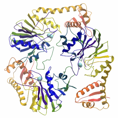
The protein crystallised in space group P21, with six monomers asymmetric unit.
Repeated attempts to solve the 2.6 A dataset by molecular replacement failed, presumably due to low homology between LACTB2 and the search models. A derivatised crystal dataset was collected and scaled to 3.2 Å. 8 mercury sites were unambiguously identified using SHELXD, with an anomalous signal extending to 6 Å. The structure was phased in a SAD experiment using SHARP. A substructure was built using PHENIX AutoBuild. This substructure was manually rebuilt and then used to rigid body refine the 2.6 Å dataset in REFMAC. Several rounds of refinement and manual rebuilding was performed using BUSTER-TNT and Coot.
The deposited structure was refined to a final resolution of 2.6 Å, R=0.179 , Rfree=0.224 and assigned the PDB code 4AD9.
|