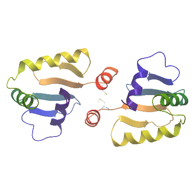
ETV1A (4AVP) Materials & Methods |
Entry clone source: MGC (Y329S & P427S mutations induced by PCR primers) |
Entry clone accession: IMAGE:30345383 |
SGC Construct ID: ETV1A-c014 |
Vector: pNIC28-Bsa4. Details [PDF ]; Sequence [ FASTA ] or [ GenBank ]. |
DNA sequence:
CATATGCACCATCATCATCATCATTC
TTCTGGTGTAGATCTGGGTACCGAGA
ACCTGTACTTCCAATCCATGGGACCC
ACATCCCAACGGCGAGGATCACTTCA
GCTCTGGCAGTTTTTGGTAGCTCTTC
TGGATGACCCTTCAAATTCTCATTTT
ATTGCCTGGACTGGTCGAGGCATGGA
ATTTAAACTGATTGAGCCTGAAGAGG
TGGCCCGACGTTGGGGCATTCAGAAA
AACAGGCCAGCTATGAACTATGATAA
ACTTAGCCGTTCACTCCGCTATTACT
ATGAGAAAGGAATTATGCAAAAGGTG
GCTGGAGAGAGATATGTCTACAAGTT
TGTGTGTGATCCAGAAGCCCTTTTCT
CCATGGCCTTTTCAGATAATTGACAG
TAAAGGTGGATACGGATCCGAA
|
Final protein sequence (His6 affinity Tag sequence in lowercase):
mhhhhhhssgvdlgtenlyfq^smGP
TSQRRGSLQLWQFLVALLDDPSNSHF
IAWTGRGMEFKLIEPEEVARRWGIQK
NRPAMNYDKLSRSLRYYYEKGIMQKV
AGERYVYKFVCDPEALFSMAFSDN
^ TEV protease recognition site
**Please note that there are two primer-induced mutations present in comparison to the MGC sequence (Y329S & P427S) - highlighted in bold and underlined above**
|
Tags and additions: Cleavable N-terminal His6 tag |
Host: BL21(DE3)-R3-pRARE2. |
Growth Medium, Induction Protocol:
For the native protein:The expression plasmid was transformed into the host strain and plated on LB-agar containing 50 µg/ml kanamycin and 34 µg/ml chloramphenicol. Several colonies were combined to inoculate a 1ml culture in TB (+ 50 µg/ml kanamycin, 34 µg/ml chloramphenicol). The culture was grown overnight, glycerol was added to 15% v/v (from a 60% stock), and the resulting glycerol stock was frozen at -80°C in 100 µl aliquots. A loopful of cells from the glycerol stock was inoculated into 20-ml of TB medium containing 50 µg/ml kanamycin and 34 µg/ml chloramphenicol and grown overnight at 37°C. 2x 1L TB medium (containing 50 µg/ml kanamycin) were each inoculated with 10 ml of the overnight culture and grown in 2.5L UltraYield baffled flasks until OD600 of 3.0. Cells were cooled to 18°C, IPTG added to 0.1mM and growth continued at 18°C overnight. The cells were collected by centrifugation then the pellets were scraped out and transferred to 50-ml Falcon tubes and frozen at -80°C.
|
Cell Extraction: Frozen cell pellets (approx 50g) were thawed briefly in a bath of warm water (20 - 37°C) then transferred to ice. Three volumes (i.e. 3 ml for every gram of cells) of lysis buffer was added. The cells were resuspended by agitating and disrupted sonication for 20 minutes on ice. Nucleic acids and cell debris were removed by adding 0.15% PEI (polyethyleneimine) from a 5% (w/v, pH 7.5) stock, stirring for 15 minutes, then centrifugation for 60 minutes at 25,000 x g.
Lysis buffer: 50 mM Na-phosphate buffer, pH 7.5, 500 mM NaCl, 10 mM imidazole, 5% glycerol, 0.5 mM TCEP, 1x Protease Inhibitors Cocktail Set VII (Calbiochem, 1/1000 dilution), and 15 units/ml Benzonase.
|
Column 1: Ni-affinity, HisTrap Crude FF, 5 ml (GE Healthcare) |
Column 1 Buffers:
Affinity buffer: 50 mM Na-phosphate buffer, pH 7.5, 500 mM NaCl, 10 mM imidazole, 5% Glycerol, 0.5 mM TCEP
Wash buffer: 50 mM Na-phosphate buffer, pH 7.5, 500 mM NaCl, 20 mM imidazole, 5% Glycerol, 0.5 mM TCEP
Elution buffer: 50 mM Na-phosphate buffer, pH 7.5, 500 mM NaCl, 300 mM imidazole, 5% Glycerol, 0.5 mM TCEP
|
Column 1 Procedure: The cell extract was loaded on the column at 4 ml/minute on an AKTA-express system (GE Healthcare). The column was washed with 10 volumes of lysis buffer, 10 volumes of wash buffer, and then eluted with elution buffer at 4 ml/min. The eluted peak of A280 was automatically collected.
|
Column 2: Gel filtration, Hiload 16/60 Superdex S75 prep grade, 120 ml (GE Healthcare) |
Column 2 Buffers:
Gel Filtration buffer: 10 mM HEPES, pH 7.5, 500 mM NaCl, 5% Glycerol, 0.5 mM TCEP
|
Column 2 Procedure: The eluted fractions from the Ni-affinity Histrap column was loaded on the gel filtration column in GF buffer at 1.2 ml/min. Eluted proteins were collected in 2-ml fractions and analyzed on SDS-PAGE
|
Enzymatic treatment and Column 3: The N-terminal His6-tag was cleaved by incubating the protein overnight with TEV protease (at 8°C). Cleaved protein was purified by passing over a 2 ml pre-equilibriated 50% Ni-IDA bead solution. Elution was done in GF buffer supplemented with step gradient of 20mM, 40mM or 60mM imidazole, with flowthorough and fractions collected. |
Column 4: Gel filtration, Hiload 16/60 Superdex S75 prep grade, 120 ml (GE Healthcare) |
Column 4 Buffers:
Gel Filtration buffer: 10 mM HEPES, pH 7.5, 500 mM NaCl, 5% Glycerol, 0.5 mM TCEP
|
Column 4 Procedure: Collected fractions from the Ni-IDA IMAC column was loaded on the gel filtration column in GF buffer at 1.2 ml/min. Eluted proteins were collected in 2-ml fractions and analyzed on SDS-PAGE.
|
Concentration: The cleaved purified protein was concentrated in a VivaSpin4 (3 K MWCO) to 16.1 mg/ml and stored at 4°C. The protein concentration was determined spectrophotometrically using e280 = 34380.
|
Mass spec characterization:
Observed mass (taking into account the primer-induced mutations) was 12421.1 (calculated mass was 12421.0 without histidine tag).
|
Crystallization: Crystals were grown by vapor diffusion at 20°C in 150nl sitting drops. A crystal was grown by mixing 50nl of protein solution (16.1mg/ml) and 100nl of precipitant consisting of 2.5M sodium formate.
Crystals were cryo-protected by bathing the crystal in mother liquor supplemented with 25% Ethylene glycol and flash-freezing in liquid nitrogen. |
Data Collection: Data collection: Diffraction data for the native dataset were collected from a single crystal at the Diamond synchrotron beamline I04, at a wavelength of 0.9795Å.
The DNA binding domain of human ETS-1 (PDB code 1GVJ) was used as a search model for molecular replacement using PHASER MR, part of the PHENIX suite. The structure was refined to an Rwork/RFree of 0.223 / 0.258 using autoBUSTER. Deposited as PDB code 4AVP.
|